Non-dangerous formations - pigment spots on the lips occur for various reasons. This may be the effect of a hereditary predisposition, or it may be a skin reaction to illness and poisoning. If dark spots occur, you should consult a doctor promptly. The earlier a problem is detected, the better the solution will be. Lip pigmentation can be either flat or in the form of a tubercle, and also has a different shade. The appearance of spots during pregnancy often goes away on its own as the woman’s hormonal levels balance. You cannot fight this disease on your own; it is important to contact trusted and specialized clinics, having previously undergone a consultation and diagnosis with a dermatologist.
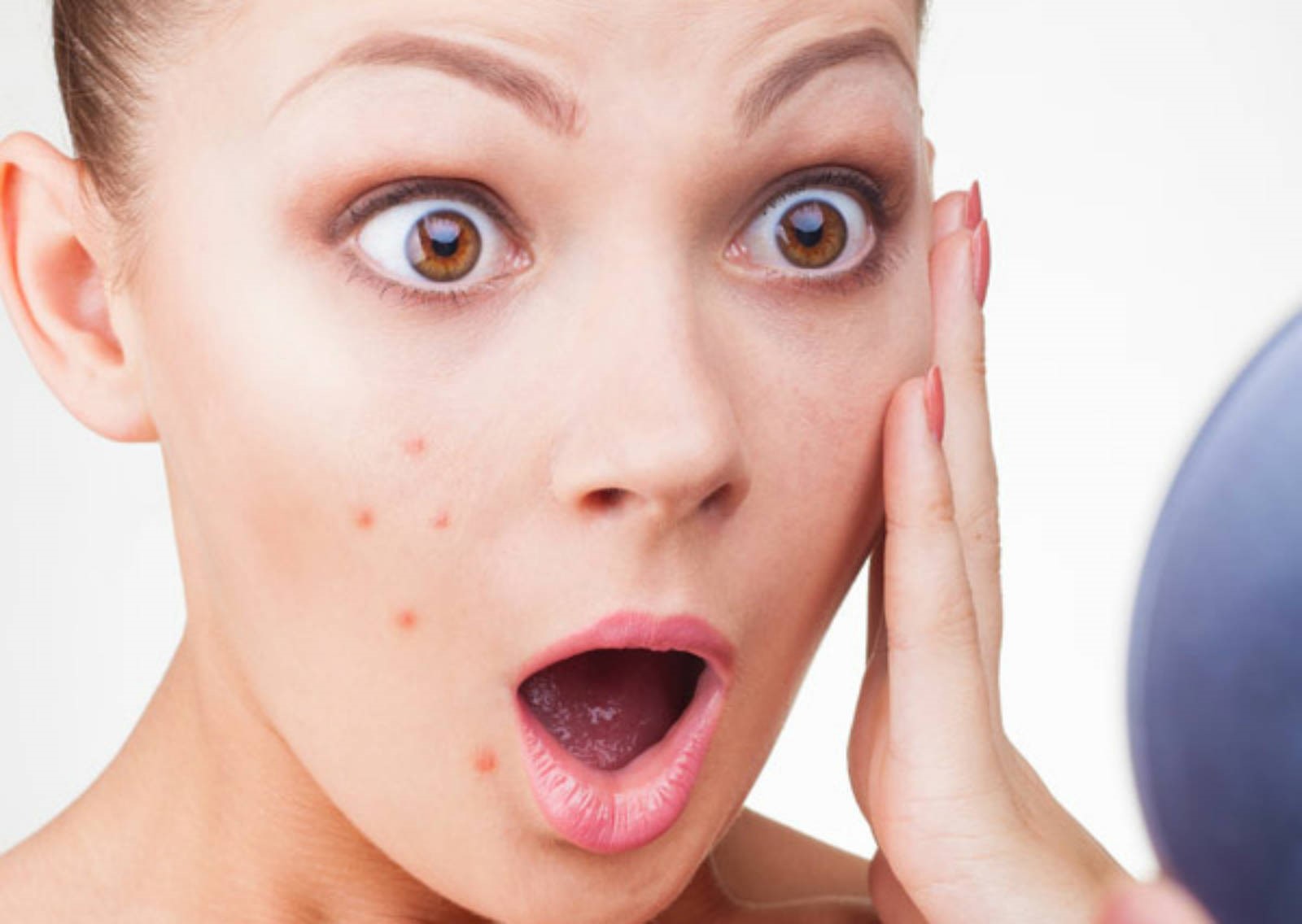
Pigment spots on the lips are a cause for particular concern in women.
What do they look like?
Pigment spots above the lip or on the lip itself are similar to freckles. They have a flat surface, a uniform tone, and appear in the form of spots of different sizes and shapes. The edges can be either smooth or jagged if lentigo (one of the types of pigmentation), which has a dense structure, appears on the lip. The color of pigmentation varies, ranging from a light yellow tint to a dark, almost black. As the lentigo grows, it can become convex and protrude like a tubercle. Any surface becomes a localization zone. Pigment spots appear above the upper lip, as well as under the lower lip or on the oral mucosa. Initially they may be barely noticeable, then darken and grow. Wide formations above the lip look like a mustache.
Reasons for appearance
Doctors do not name the exact nature of the disease. Characteristic factors influencing the manifestations of pigmentation in the lip area are:
- exposure to ultraviolet rays;
- reaction to low-quality decorative cosmetics;
- injury to the surface of the lips;
- hormonal disbalance;
- hereditary factor;
- Peuttz-Jeghers syndrome;
- stress and depression;
- low immunity;
- infestation by poisons or poisoning;
- reaction to synthetic drugs and medications.
 Stress, sun rays, cosmetics, injuries, and a deficiency of nutrients in the body contribute to the appearance of pigment spots on the lips.
 There are many guesses and arguments about the reasons for the development of pigmentation on the lips. Diagnostics helps to reveal a more accurate picture of the development of tumors. The appearance of pigmentation on the face is often associated with external discomfort, especially in women. The reasons for the appearance also depend on the type and type of spot. Below is a table with characteristic pigmentation on human lips.
Pigmentation on the lips often becomes a reflection of serious diseases. In such cases, urgent treatment by a doctor is necessary.
Diagnosis of the disease
 Treatment for age spots on the lips is prescribed by a doctor after diagnosis.
 Removal of pigment spots does not take place without preliminary diagnosis. Conducted by a dermatologist, starting with a visual examination of the patient for symptoms. This problem is easy to recognize, but to clarify the picture, techniques using special equipment are used. Dermatoscopy is performed, which on an enlarged scale indicates the pathology of the development of the disease, spread to nearby organs and tissues and the depth of its germination. Sometimes computer diagnostics are prescribed. This method is based on studying a photograph of the formation previously taken by the camera of the device. A blood test is taken to determine whether the lump is benign. Tumor markers are used to determine the presence of cancer cells.
Treatment of the problem
Every woman who has had this problem dreams of getting rid of a pigmented mustache. Since this is a harmless neoplasm, treatment is not necessary. You need to fight not with the stains on the lips themselves, but with the nature of their occurrence. It is impossible to remove seals on your own; such attempts to remove spots are fraught with complications and can lead to a number of dermatological problems. If the problem is related to a deficiency of immunity, by increasing it, the spots will go away on their own. Vitamin complexes and beneficial microelements can help remove stains. Nodules are removed by surgery only in urgent cases and in the presence of a malignant tumor. After the operation, a scar remains. The excised material is sent for histological analysis.
In cosmetology and dermatology offices, laser destruction and pigmentation removal using radio or electric waves are used. They penetrate deep into the tissues, reducing pigmentation. The procedures are painless and leave no traces. There is no rehabilitation course. A person can immediately begin household chores immediately after the procedure. The number of receptions in each case individually depends on the degree of manifestation. Conservative treatment methods include whitening masks, creams and tonics. They reduce the color intensity, but do not completely remove pigmentation. Treatment is used only after consultation with a dermatologist and appropriate diagnosis.
Appearance any pigmentation on the lips should not be ignored. If a blue dot appears on the lip, even more so. Because it always indicates the presence disruptions in the functioning of the body.
Interesting! Doctors distinguish two main types of spots - youthful and senile. Both cases have their own characteristics and causes. Before starting treatment for a defect, you should understand what caused it to appear.
Causes of dots on lips
Spots and dots on the lips can vary in size, shape and color. However, special attention should be paid to blue, intensely pigmented dots, since they most often appear as a consequence of serious illnesses:
Only a doctor can make a correct diagnosis and prescribe treatment in each specific case.
Spots caused by harmful effects of ultraviolet rays, are successfully eliminated using bleaching agents and appropriate vitamin complexes.
If the spots are intensely colored, injections of ascorbic acid help well. Appearance large quantity blue dots on the lips (hyperpigmentation) requires oral administration of folic acid, aevit and riboflavin.
If any other diseases are discovered during research, the intervention of specialists in the required field is required - oncologists, gastroenterologists, dermatologists. Treatment in this case will be comprehensive, and as your overall health improves blue spots will disappear on their own.
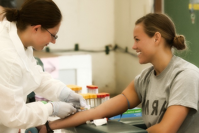 Photo 2: Treatment of any pigmentation on the lips depends on the causes of the defect, so first of all you will need to undergo an examination and a blood test. Source: flickr (Dave Black)
 Homeopathic medicines
The success of homeopathic treatment, first of all, lies in in an individual approach to every patient. The homeopathic doctor prescribes a remedy suitable constitutional type of the patient. In this case, the chosen remedy will affect improvement of the body as a whole.
Depending on the type of stain on the lip and the patient’s psychotype, the following drugs may be prescribed:
- Arnica (Arnica montana). The drug promotes the resorption of compactions and is used to treat warts and venous nodules on the lips. The constitutional type of Arnica is full-blooded, good-natured people. Most often they are friendly, but during illness they become moody and irritable.
- Calcarea fluorica. Effectively fights vascular tumors, increases the tone of capillaries and blood vessels, and helps in case of helminthic infestation. Prescribed to patients with malocclusion and severe asymmetry of the bone skeleton.
- Silicea. The product effectively fights papillomas and helps eliminate hyperpigmentation on the lips. Psychotype - thin, sickly people who tend to get nervous over trifles. They often freeze and do not tolerate mental stress well.
- Phosphorus (Phosphorus). The drug is prescribed if the appearance of defects on the lips is caused by dysfunction of the liver and adrenal glands. Constitutional type of the drug - tall, stooped people with soft blond hair. The character is sensitive, touchy and vulnerable.
- Bellis perennis. The drug fights the manifestations of excessive pigmentation on the lips and has whitening properties. Most often prescribed to older people who complain of constant fatigue and memory problems.
The popularity of homeopathic treatment is primarily due to proven effectiveness of drugs. In addition, the absence of side effects that often occur when using traditional medications can be considered a big plus. Any drug prescribed individually by a homeopathic doctor, therefore, when a blue dot appears on the lip, consultation with a specialist will be the first step towards successful treatment.
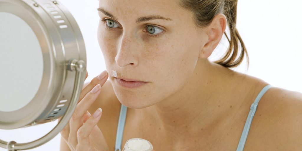
The effect of ultraviolet radiation on the body has both positive and negative effects.
When we go to the beach, we protect our body with sunscreen, our eyes - sunglasses, and the head with a hat, but we often forget that lips also need special care.
Lip pigmentation: looking for a cause-and-effect relationship
Sudden pigmentation on the lips is often a consequence of using lip gloss in the summer. Glitter directs and concentrates ultraviolet rays, which is the cause of spot pigmentation. To avoid this, during periods of increased insolation, do not use glossy glosses, giving preference to hygienic lipstick. More tips You will find in our material Proper care behind your lips in the heat. 5 simple tips.
Pigmentation on the lips should be taken very seriously, since in most cases it can be an indicator of serious diseases. For example, the most common cause of age spots on the lips is chloasma. Basically, this cosmetic defect bothers women only during pregnancy and disappears on its own after childbirth. But if a woman is not pregnant, the appearance of chloasma may indicate a malfunction of the ovaries, adrenal glands or liver.
In addition, spots in the lip area may be the result of progressive degeneration of benign or malignant cells of the mucous membrane.
If newly appeared spots on the lips are small in size, these may be seborrheic cysts (Fordyce granules).
The combination of pigmentation of the mucous membranes and skin may indicate a more serious disease called Peutz-Eigers syndrome. Associated changes are polypous changes in the mucous membrane of the stomach and small intestine.
How to get rid of lip pigmentation and make lips flawless
A prerequisite is to determine the reason why these spots arose. To do this, you should consult a dermatologist.
If lip pigmentation is not a manifestation of any multisystem disease, you may be prescribed the following treatment:
- Taking vitamin complexes. You may be prescribed ascorbic acid, for greater effectiveness - by injection. Also good result indicates taking folic acid preparations (Vitamin B9), preparations containing a compound of vitamins A and E and riboflavin.
- Procedures. Among hardware procedures, laser coagulation, which is considered the most modern method, can help.
- Whitening creams. At night, a special serum is applied to the affected area to brighten the skin of the face and lips. It can only be applied to problem areas.
Smoking can cause a characteristic discoloration of the mucous membrane, characterized as smoker's melanosis. This condition is caused by pathological deposition of melanin in the cells of the basal layer of the mucosal epithelium. The relationship between melanosis in smokers and inflammatory changes caused by high temperature, inhalation of tobacco smoke and absorption of exogenous pigments has not been established.
Melanosis develops in older people, heavy smokers. It appears as a diffuse brown spot, reaching several centimeters in size. Most often, melanosis is localized on the gum of the anterior part of the alveolar arch of the lower jaw and the mucous membrane of the cheeks, less often on the lips, palate, tongue, and floor of the mouth. The degree of pigmentation ranges from light to dark brown and depends on the intensity of smoking. Usually, dark brown areas are observed on a light brown spot with unclear boundaries. Melanosis in smokers is usually accompanied by brown staining of the teeth and halitosis ( bad smell from mouth). Smokers' melanosis in itself is not a precancerous condition, but patients with this lesion should be monitored, given the high risk of developing other, more serious changes in the oral mucosa.
Melanotic spot (focal melanosis).
Melanotic spot- an area of pigmentation with clear boundaries, localized on the lip or in other parts of the oral cavity and caused by increased local deposition of melanin in the basal cells of the epithelium. The melanotic spot is usually single, does not exceed 1 cm in diameter, does not manifest clinically and is usually observed in fair-skinned individuals aged 25 to 45 years. The cause of the formation of a melanotic spot appears to be trauma or an inflammatory process. Typically, a melanotic spot is localized in the middle of the lower lip, less often - on the gum, mucous membrane of the cheek, and palate. The color of the spot is uniform and can be blue, gray, brown or black. Patients with this lesion should be examined periodically and, if changes occur, undergo a biopsy.
Nevus of the oral cavity.
Nevus- damage to the mucous membranes and skin, consists of an accumulation of nevus cells with high content melanocytes in the epithelium or dermis. Typically, a nevus is localized on the skin, but sometimes it is also detected in the oral cavity. Nevi are divided into two large groups - congenital and acquired. Congenital nevi are detected at birth; they are otherwise called birthmarks. Compared to acquired nevi, they are larger in size and more often undergo malignant transformation.
Acquired nevi, or moles, appear in more late dates life and have the appearance of dark, slightly protruding papules or hemispherical nodules above the skin. Depending on the pigment content in them, they can be brown, gray or black. Sometimes nevi do not contain melanin and have pink color. Nevi in the oral cavity are rare, mainly in women. They usually take the form of a small pigmented spot or nodule with clear boundaries, localized on the palate or buccal mucosa and not causing painful sensations. After puberty, the size of nevi usually does not change.
Benign nevi are divided into four subtypes depending on the histological picture and clusters of nevus cells. Most often found in the oral cavity is an intramucosal nevus, consisting only of ovoid nevus cells located in the connective tissue. This nevus resembles an intradermal nevus and appears as a dark, raised papule on the skin from which hair often grows. However, with intramucosal nevus, hair growth is rarely observed. An intramucosal nevus usually protrudes above the level of the mucous membrane and has Brown color and diameter from 0.4 to 0.8 cm.
The second most common nevus of the oral cavity- blue nevus. It gets its name from its characteristic blue or blue-black color, which is caused by spindle-shaped nevus cells located deep in the connective tissue. These cells arise in embryogenesis from the neural crest and, for some reason, do not reach melanocyte maturity. Typically, this nevus appears as a small blue spot, the color of which fades with age. The palate is the most common location for blue nevus. Cases of malignant transformation of blue nevus of the oral cavity have not been described.
Complex nevus of the oral cavity, as the name suggests, consists of nevus cells located both in the epithelium and in the lamina propria of the mucous membrane. Complex nevus rarely becomes malignant. Borderline nevus is a type of acquired nevus; its cells are localized in the epithelial layer bordering the lamina propria. This is the rarest type of nevus.
Borderline nevus usually does not protrude above the surrounding surface, is brown in color, less than 1 cm in diameter and is usually localized on the mucous membrane of the palate and cheeks.
Sometimes on the mucous membrane of the oral cavity Hutchinson's melanotic freckles(malignant lentigo). Melanotic freckles are usually observed on the face in patients over 50 years of age. They have the appearance of grayish-brown spots of irregular shape, are capable of infiltrating growth within the plane in which they are located, and undergo malignant transformation.
Nevi difficult to distinguish from malignant pigmented lesions, so a biopsy should be performed in all cases to confirm the diagnosis.
Blue mole on the lip may be a sign of a venous lake (see photo). A venous lake is an enlarged vein in sun-damaged skin that appears as a small, dark, blue-purple papule that is capable of discoloration.
Venous lakes are common on sun-damaged skin of elderly patients. Venous lakes occur as a result of acquired skin damage due to sun exposure and subsequent loss of skin elasticity (solar elastosis).
Clinical picture
Very often, patients with venous lakes worry about a possible malignant disease. Venous lakes with blood clots can be painful. The lesions resemble varicose veins on sun-damaged skin.
Venous lakes are slightly raised, dome-shaped dark blue lesions measuring 0.2-1.0 cm in size, which consist of an enlarged, blood-filled vascular canal. Multiple lesions may be present.
The venous lake is an asymptomatic soft dark blue-violet papule with a diameter of 2-10 mm, which discolors when pressed. Multiple lesions may be present on the mucous membrane of the lips, especially on the lower lateral red border of the lips. Lesions can also be on the ears.
Raised lesions and lesions exposed to trauma (eg, labial mucosa) may be itchy and painful, suggesting thrombosis. Injured lesions bleed easily and form a hemorrhagic crust.
Diagnostics
A skin biopsy shows dilation of the thin walls of the venules located in the upper layers of the skin or under the mucous membrane.
Venous lakes may resemble pigmented lesions such as blue nevi and malignant melanoma; venous lakes are completely discolored during diascopy.
Injured venous lakes can become crusty and look like herpes labialis.
Patients with HIV-associated Kaposi's sarcoma develop multiple blue-violet nodules on the mucous membranes that resemble venous lakes.
Venous lakes persist and may increase in size over time.
Lesions that are often traumatic or cosmetically unpleasant, as well as lesions that interfere with eating and speaking, should be treated, although they tend to recur. After local or regional anesthesia, the venous lake is opened and cauterized. Lasers are also effective for removing vein lakes. If the lesions change quickly, you should contact a dermatologist.
T.P.Hebif
"Blue mole on lip" and other articles from the section