“Knowledge is power” - we got used to this term from our Soviet childhood, and we don’t think that knowledge is life. First of all, it concerns breast cancer. The average life expectancy in patients with newly diagnosed breast cancer is 12-15 years in Western Europe and 3-5 years in our country. Such a significant difference in the outcome of the disease, given the generally common approaches to the diagnosis and treatment of the disease itself, was analyzed in detail and the results depressed the experts.
The first fundamental difference is the woman's careful attitude to her own health, timely assessment of the early symptoms of the disease. correct perception of the need for regular check-ups. As a result, over 90% of European women and only 30% of our compatriots seek medical help for the first time at the 1st stage of breast cancer. Many are simply afraid to come to the doctor's office so as not to hear the diagnosis of cancer. Most of our women seek medical help for the first time already at the 2nd and 3rd stages of cancer. In many ways, this difference is dictated by the low level of knowledge and desire to protect their own health. Then the psychological perception of the diagnosis "cancer" as a sentence that no longer gives a chance for healing.
The second fundamental difference between our patients is the fact that after the diagnosis of "cancer" is made, about 90% of all patients during the first six months, the most valuable for them, practically disappear from the doctors' field of vision, trying to achieve treatment with "folk remedies".
Given that every 8th woman develops breast cancer during her life, it is necessary to firmly grasp the main early manifestations of cancer, the principles of self-diagnosis and early diagnosis of breast cancer.
Who needs to be particularly vigilant and alert about breast cancer?
The main risk factors for developing breast cancer are caused by disturbances in the hormonal balance of a woman. First of all, this is a burdened family history (breast cancer along the female line - in a sister, mother, grandmother), changes in the mammary glands (after injuries, childbirth; fibrocystic mastopathy), early menopause (especially before 30 years of age as a result of surgical castration, for example, bilateral resection of the ovaries after apoplexy), late childbirth or childlessness over the age of 30 years.
How does breast cancer manifest itself?
Warning signs for alarm can be symptoms such as:
- "bump" or seal in the area mammary gland, not disappearing after menstruation;
- focal changes in the contours, size or shape of the breast, mainly on one side;
- discharge from the nipples (light liquid or bloody);
- a bump or lump in the breast the size of a pea;
- redness of the nipple or skin of the breast, retraction of the nipple on one side;
- stone-like compaction in the breast;
- change in the appearance of the nipple or breast skin (inflammation, peeling, dimpling, or wrinkled skin);
- the area on the chest is distinctly different;
- enlarged lymph nodes under the arm;
- swelling of the tissues of the armpit and shoulder.
These changes can be seen first only by women themselves. It is necessary to learn not only to carefully see the changes in your body, but also to master the practical skills of self-examination, including self-palpation of the breast and nearby lymph nodes.
Breast self-exams should be done monthly, approximately 3-5 days after your period. It must be remembered that more than half of women find changes in the mammary glands, and only one in eight gets breast cancer.
If you suspect this insidious disease, you should urgently seek help from specialists (mammologist and oncologist). In a medical institution, an examination and professional palpation will be performed mammary glands and surrounding tissues, a detailed survey was conducted to clarify the history of life and disease (history taking), and adequate diagnostic methods were prescribed, based on the results of which it will already be possible to judge the presence of the disease and its stage.
The following tests help detect breast cancer:
- ultrasound - ultrasound;
- cytological examination of a smear separated from the nipple of the mammary gland;
- mammography (X-ray);
- magnetic resonance imaging - MRI.
- biopsy of a suspicious tissue area under ultrasound control.
In some cases, especially with nodes less than 5 mm, a precision computerized biopsy can be performed using an automated system.
The material obtained from the biopsy must necessarily undergo a histological examination, preferably in two different centers, as well as with a positive diagnosis of cancer - an immunohistochemical study and a study on the expression of estrogen, progesterone and other receptors by tumor cells.
As necessary, the doctor may prescribe additional studies that help assess the general condition of the body, the prevalence of the tumor process, determine the presence of metastases in the lymph nodes or other organs, and identify comorbidities:
- radiography of the lungs;
- Ultrasound of organs abdominal cavity and small pelvis;
- general clinical tests, examinations;
- biopsy of peripheral lymph nodes;
- skeletal scintigraphy;
- CT chest and abdominal organs.
In the presence of what factors is breast cancer manifested?
- age 40 and older;
- in the blood - an increased level of estrogen;
- long-term use of high-dose hormonal drugs;
- 1st line relatives who have been diagnosed with breast cancer;
- previous oncological disease of the breast or ovary;
- the first pregnancy was at the age of 30 or older, or the woman is infertile;
- prolonged contact with radioactive isotopes and/or hard X-ray sources;
- absence of pregnancy and childbirth;
- atypical changes in the epithelium of the thoracic ducts (epithelial hyperplasia) - are detected during a cytological examination of the smear from the mammary gland;
- the onset of menstruation before the age of 12 and / or the onset of menopause later than usual;
- metabolic and endocrine disorders (obesity, type 2 diabetes mellitus);
- excessive consumption of fatty foods.
What is breast cancer prevention?
First of all, the development of this disease can be prevented by observing the normal physiological rhythm of life (pregnancy, childbirth, prolonged feeding) with the exclusion or reduction to a minimum of the number of abortions. It is also important for patients to treat precancerous seals in the mammary glands in a timely manner.
Monthly breast self-exams should be performed. Women over 40 years of age undergo annual preventive examinations and once every 2 years - mammography. An annual mammogram is recommended for women at risk (regardless of age) and over 50 years of age.
Neoplasms that arise in the mammary gland may have malignant form, differ in aggressive cell proliferation leading to metastases.
Symptoms of breast cancer
How to determine the glands in women, what symptoms should be alarming? There are many signs of breast disease, but little attention is paid to them. This is due to the fact that they do not cause discomfort in the early stages of pathology. Meanwhile, time is the main factor in the treatment of breast cancer.
Important! 80% of women were able to avoid death due to a timely diagnosis of breast cancer and timely therapy.
Symptoms of breast cancer are different, it depends on the shape and size of the anomaly, the degree of spread and location.
Common symptoms of breast cancer:
- The presence of seals;
- discharge from the nipple;
- the nipple sunk in.
Unusual signs of breast cancer:
- Constant feeling of pain in the back;
- fleeting asymmetry of the bust;
- peeling, irritation, redness, itching appeared on the skin of the chest.
Breast cancer and its symptoms in each form of the disease must be considered in more detail:
- With a nodular form, a neoplasm can be recognized by a solid ball. Its diameter can be from 0.5 - 5 cm or more. With this type of disease, all of the following symptoms will also appear.
- Diffuse breast cancer is divided into three types:
- Armored - with this type of pathology, a malignant neoplasm spreads through the gland in the form of a "crust", which tightens and reduces the size of the affected breast.
- Erysipelatous - the skin on the surface of the bust becomes red, there is a feeling of pain, body temperature may rise to 40 ° C.
- Pseudo-inflammatory - signs, as with an erysipelas-like form of the disease. Because of this symptomatology, it is difficult to correctly diagnose the pathology, the patient is prescribed therapy for those diseases that are present in the name of this form of oncology.
All three of these species are very aggressive. Cancer cell growth occurs at lightning speed and spreads throughout the bust, without clear boundaries.
There are cases when the neoplasm manifests with metastases to the lymph nodes on the affected side. In this case, the tumor is not detected, it is difficult to diagnose cancer. In such cases, this type of pathology is called "hidden oncology".
If the formation is insignificant in size, then no obvious manifestations are observed. On her own, a woman can feel such a tumor only with a small size.
A malignant node, as a rule, is motionless during palpation, and if displacement occurs, then it is insignificant, does not cause pain, has an uneven surface and stone density.

In oncology, the skin over the neoplasm is significantly different - it becomes wrinkled, folds appear. Retracts, becomes edematous. There is a sign of "lemon peel". In rare cases, there is a "cauliflower" - the germination of the tumor outward, through the dermis.
Important! If such signs were revealed during self-diagnosis, then the condition of the lymph nodes should be checked. You should not be concerned about their slight increase, the feeling of pain and their mobility during palpation. But in the case when the lymph node is large, merged into one whole with others and dense in consistency, then this is the place of localization of metastases.
One of the symptoms of oncology in this part of the body can be swelling of the hand on the side of the neoplasm. If there is such a recognized sign, then you should know that these are already the last stages of the disease. Metastases penetrated into the axillary lymph nodes and sealed the outflow of fluid and blood from the arm.
To summarize, all that was listed above, it is worth indicating the main symptoms of this disease:
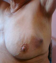
- Any change in shape - a decrease or increase in the size of one of them, a sinking or displacement of the nipple.
- Change in the dermis mamma - sores in the area of the areola, nipple. Change in color anywhere on the skin - blue, red, or yellow. Thickening or contraction of a limited area - "lemon peel".
- A hard, immovable knot.
- In the axillary zone, lymph nodes are enlarged, giving slight pain when palpated;
- When pressing on the nipples, discharge appears - with ichor or transparent;
Important! It is impossible to detect breast cancer on your own in the early stages. Therefore, it is important to visit a mammologist regularly.
Of course, there are much more signs of oncology of this anomaly, but it is the women listed that are most often noted.
How to identify the pathology of the disease at home is of interest to many women.
self-examination
Experts recommend that all women who are at risk of developing cancer have regular self-examinations. Diagnostics should be done at the same time of critical days. This is due to the fact that during the menstrual cycle, the female breast undergoes changes in the structure and size of mamma.
A more suitable time for examination is on the fifth, sixth day from the beginning of the cycle. In menopause, the procedure should be carried out by choosing one constant day in the month.
The study consists of six stages, which include:
- Inspection of underwear - this is due to the fact that with changes occurring in the mammary glands, discharge may appear. They may be invisible on the nipple, but leave a mark on the bra in the form of ichor, caked pus, greenish or brown spots.
- The appearance of the bust - for a visual inspection, you need to stand in front of the mirror and undress to the waist and carefully examine the mamma separately. First of all, it is necessary to pay attention to the symmetry of both breasts - they should be located on the same level, move evenly with arms raised or wound behind the head, with the turns and tilts of the torso. During this check, attention should be paid to whether or not one of the breasts is shifted or fixed to the side. Next, raise your arms above your head and carefully examine the bust for an example of the displacement of the latter up, down or sideways.
With such a check, you need to look to see if changes appear in the form of dents, bulges, nipple retractions and whether liquid starts to stand out from it at the time of these movements.
- The general condition of the dermis of the bust - it is necessary to pay attention to elasticity, skin color. Does it have redness, diaper rash, rash, "lemon peel", sores.
- Standing palpation - this procedure can be carried out in the shower. With a soapy hand, mamma can be easily felt. The left breast is examined right hand and vice versa. Palpation is carried out with the help of fingertips, and not their tips. Connect three or four fingers and make penetrating movements in a spiral. If the breast is large, then it should be supported by the hand during the examination.
The first stage of such a check is called superficial - the pads do not penetrate deep inside. In this way, formations under the skin can be detected.
After that, you can proceed to the second stage - deeper palpation. In this form, the fingers penetrate gradually to the very ribs. This examination is carried out in the direction from the collarbone to the rib and from the middle of the sternum to the armpit inclusive.
- Probing lying down is an important step in the self-diagnosis of breast cancer. This is due to the fact that in this position the gland is well palpated. For the study, it is necessary to lie on a hard surface, placing a roller under the chest area. One limb should be extended along the body or placed behind the head. In this position, diagnostics can be carried out in two ways - square and spiral.
Square - mentally divide the entire chest part into squares and feel each section from top to bottom;
Spiral - from the area of \u200b\u200bthe armpits to the nipple, use the fingertips to move in a circle.
- An examination of the nipple is necessary in order to timely determine breast cancer. After all, cancer detected in the early stages is easier to treat.
When examining the nipple, you should pay attention to its shape, color - whether they have changed. Have cracks and ulcers appeared? The area around the nipple and the nipple itself must be palpated in order to exclude the appearance of a tumor.
At the end of the self-examination, take the nipple with two fingers and press on it. This is necessary in order to find out if there is a discharge from the nipple.
If after the last diagnosis at home there has been a certain shift in the negative direction, then you urgently need to contact a specialist who, after conducting a full study, taking into account the clinical symptoms, and the treatment will be able to prescribe the appropriate diagnosis.
Important! Before panicking when you find seals in your chest, you should consult a specialist. After all, only he can say it is a carcinoma or adenoma of the mammary gland, the symptoms of which during self-examination may resemble a malignant tumor.
This disease occupies the first place in frequency among women, the second - after among the male and female audience, since cancer also occurs in men (less than 1%).
What is breast, mammary gland, breast cancer?
The sweat gland that evolved into the mammary gland is called the breast. The structure of the female and male mammary glands is identical, but the degree of their development is different. During puberty, against the background of hormonal changes, the development and functioning of the breasts of boys and girls begins to differ, since in boys the body starts processes that are different from the female internal processes.
With breast growth that begins before the onset of menstruation, the girl turns into a woman, which indicates that the breast is a hormone-dependent organ.
It is important to know! Since the breast consists of a right and left organ, hormonal changes affect both breasts in the same way.
Therefore, with the ongoing changes in the chest, you can correctly respond to the current process. For example, if both breasts ache before menstruation, then this is due to premenstrual swelling of the gland. But with pain in only one breast, you should immediately consult a gynecologist-mammologist, if it is not associated with scuffs from the bra. Pain can be associated with pathological processes inside the breast, like breast cancer.
Breast anatomy
The pectoral muscle holds both mammary glands, which are based on glandular and adipose tissue. The size of the breast depends on the amount of adipose and glandular tissue. The connective tissue divides the gland into 15-20 lobes, and each lobe into many small lobules with a diameter of 0.05-0.07 mm, the space between which is filled with fatty tissue. At the site of attachment of the gland to the chest wall, there is also adipose tissue in the form of a pillow. It supports the gland and creates the shape of the breast.
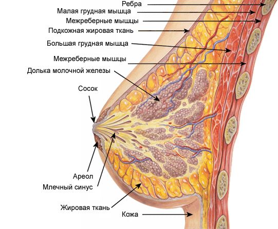
Separate mammary glands, consisting of many tubes with extensions at the end - alveoli (microscopic vesicles) make up the glandular part, located in the lobules of the gland. Milk is produced in the alveoli. The excretory ducts (tubules) transport it to the gland through the terminal sections of the tubules and further dilated lactiferous sinuses that open on the nipple. The nipple is located just below the center of the chest and opposite the 4-5 rib gap. The shape of the nipple is conical in nulliparous women and cylindrical in those who have given birth.
On the surface of the nipples and their areolas (pigmented area with a diameter of 3-5 cm) there are muscle cells with a large number of nerve endings, due to irritation of which, when fed from the nipples, breast milk. The color of the nipple and areola is pinkish or dark red in nulliparous women, brownish in those who have given birth.
Nipples with nerve endings become a sensitive erogenous zone and rise with the contraction of muscle cells during sexual arousal (erection). Small vestigial mammary glands of Montgomery are also present in the circle near the nipples in the form of small elevations.
The nipples are covered with wrinkled skin with small holes at the top - milky pores (ends of the milk ducts) with a diameter of 1.7-2.3 mm. When some milk ducts merge with each other, the number of holes reaches 8-15, which is less than the total number of ducts.
The mammary glands are supplied with blood through the thoracic arteries: internal and lateral.
Type of mammary gland at maturity
Until the age of 11-12, the mammary glands in girls consist of mammary glands in the form of short tubes without branching and alveoli. Against the background of estrogen, which is produced by the ovaries, the growth of the milky tubes in length begins, and at their ends - the alveoli with a simultaneous increase in the amount of connective, adipose and glandular tissue. This is how a woman's breast size is formed.
The appearance of the breast during the menstrual cycle
During menstruation, cycles of changes in the mammary gland begin. In the second phase of the cycle, progesterone promotes the development of alveoli within 12-14 days. When the production of progesterone stops, the alveoli stop developing and disappear until the next cycle begins.
At the end of the cycle of menstruation, the size of the mammary glands slightly increases, they “sharp” with simultaneous discomfort and soreness. This is how premenstrual syndrome begins.
Type of breast during pregnancy and after childbirth
Pregnancy contributes to the full development of the mammary gland, since there is a long-term release of progesterone, which activates the development of the alveoli. By the end of pregnancy, another hormone is produced - prolactin, which contributes to the production of colostrum in the alveoli - a special secret that contains a lot of protein and less lipids, unlike breast milk.
The synthesis of the hormone prolactin, which is responsible for the secretion of milk and stimulation of the development of milk lobules, occurs in the pituitary gland. Men also produce prolactin. Elevated prolactin levels lead to stress and problems in the breast.
After childbirth, the mammary gland produces breast milk - lactation begins under the influence of the alveoli of the main hormone - oxytocin, as well as thyroid hormones.
So, the functioning of the breast is affected by: progesterone, prolactin and oxytocin, as well as insulin, so women with diabetes are more likely to get breast cancer. The state of the thyroid gland is directly related to the mammary glands and uterus.
Under the influence of hormones produced by the thyroid gland: thyroxine (T4) and triiodothyronine (T3) occurs:
- regulation of metabolism in the body;
- cardiovascular activity;
- work of the gastrointestinal tract;
- functional work of the reproductive system;
- mental activity.
Interesting to know! The left breast is larger than the right breast. Disorder of the endocrine system leads to an increase in men's breasts and secretion of milk. In newborns, the mammary glands are capable of pathological secretion, the so-called "witch's milk".
The development of the mammary glands may be abnormal, so observe:
- amastia - unilateral or bilateral atrophy of the mammary glands (MF);
- macromastia - an increase in the breast up to 30 kg on both sides;
- polymastia - the presence of additional MF in the area of \u200b\u200bthe armpits;
- polythelia - abnormal development of the breast in the form of several nipples along the line of the body.
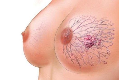
Breast cancer - what is it?
An epithelial tumor originating from the lobules or ducts of the gland is called breast or breast cancer. Most often, malignant oncopathology occurs - with late diagnosis and with a negative outcome.
Breast cancer (BC) can be triggered by the following factors:
- high blood levels of estrogen;
- taking hormonal contraceptives;
- drugs with hormones that regulate the menstrual cycle;
- the use of hormone replacement therapy in menopause;
- the presence of relatives in the 1st female line with breast cancer;
- first pregnancy after 30 years;
- infertility;
- over 40 years old;
- previous ovarian cancer or MOH;
- contact with a radioactive source;
- the occurrence of changes in the breast, such as atypical epithelial hyperplasia;
- endocrinological and metabolic disorders - thyroid disease, obesity;
- increased consumption of fatty foods;
- early onset of menstruation (at 9-11 years old);
- late onset of menopause.
With an increase in the size of the breast, the risk of developing cancer increases.
Causes of tumors, precancerous diseases of the breast
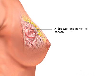
Cancer can develop in connection with previous pathological processes in the tissues of the breast - repeated dyshormonal hyperplasia, in which foci of fibrocystic mastopathy (fibroadenomatosis) are formed.
Endocrine disorders against the background of ovarian diseases, improper feeding of the child, in connection with abortions become the causes of these pathological processes.
The causes of breast cancer in women may be due to mutations occurring in healthy breast cells. Exposure to carcinogens, as well as cancer risk factors, can change DNA, which is why mutations and the transformation of normal cells into oncogenic ones occur, especially with their frequent division.
A malignant tumor in the breast can develop due to the presence of:
- mechanical injuries: bruises of the breast with hematomas, bruises;
- elevated estrogen levels;
- disorders of the adrenal glands and other endocrine glands;
- frequent abortions, which excludes lactation;
- bad habits: smoking, increased consumption of animal fats and beer;
- daily stress, sedentary lifestyle;
- in men - a concomitant disease - gynecomastia.
Frequent precancerous diseases:
- fibrocystic mastopathy is characterized by benign hormonal and morphological changes in breast tissue;
- mastitis - refers to purulent inflammation of the breast, which often occurs after childbirth during the formation of seals due to a sharp excess of milk;
- skin lesions of the breast without tumors combine eczema of the nipple, candidiasis of the folds under the breast, bacterial infections.
Breast cancer: symptoms and signs of the disease in women and men
Signs of breast cancer in the early stages of a woman may not be noticed during a thorough self-palpation of the breast. Even experienced specialists may not detect a tiny tumor by palpation. You can determine all changes in the breast using mammography. With certain risk factors, the diagnosis is confirmed by screening for ultrasound or MRI.
If signs of breast cancer in the form of a tumor began to be detected during palpation at home or at a doctor's appointment, then this already indicates the development of a more serious stage of cancer.

During daily breast examinations, you should be alert if you have:
- redness and peeling of the skin;
- visual change in the nipple and pain in it;
- discharge from the nipple;
- a lump or small lump, especially in one breast;
- deformation and swelling of the breast;
- changes in the contour of the breast during palpation, which is called a symptom of the site;
- "lemon peel" - noticeable pores on the skin;
- sores on the skin;
- retraction of the nipple and over the tumor - the skin;
- enlarged lymph nodes under the armpits.
If breast cancer is suspected, the symptoms can be checked with diagnostic tests: biopsy and mammography, which will show the tumor even through the dense tissue of the breast.
Do breasts hurt with cancer? Answering this question, we can add that a pulling pain appears not only in the chest, but also in the back between the shoulder blades during a night's sleep. At the same time, deep breathing and / or body position are not associated with it.
Symptoms and signs are most often manifested in adverse environmental conditions, negative impact harmful chemicals in production and from household chemicals, penetrating radiation, solar radiation, widespread and unreasonable use of drugs by residents of large industrial cities.
Breast cancer in men (teenagers and the elderly) can occur if:
- gynecomastia - an increase in breast tissue in violation of the balance of hormones;
- the appearance of a tumor or liver disease, which leads to increased production of estrogen - the female sex hormone;
- the use of certain drugs in the treatment of peptic ulcers and diseases of the heart and blood vessels that cause gynecomastia;
- Klinfelter syndrome, a rare genetic disease that causes gynecomastia and increases the risk of breast cancer.
![]()
Risk factors for getting sick also include heredity, radiation exposure, physical inactivity and obesity. Symptoms clearly indicate breast cancer in men, which is characterized by neoplasms in the breast located under the nipple or in the areola area. A bloody substance will come out of the nipple. In the last stages of cancer, they will be disturbed by: ulcerations of the skin, a rapid increase in axillary lymph nodes and their compaction. In this case, cancer can spread beyond the breast, since in men it is smaller than in women. The prognosis for recovery can be disappointing.
Other symptoms of breast cancer
When examining and suspecting cancer, the doctor pays attention to the nature of the seals, which are then examined in the laboratory. Oncology is indicated by nodes (single or group) with clear contours, painless, with a dense texture, limited mobility and the presence of wrinkled skin retractions above the node (nodes). In this case, lymph nodes can be palpated under the armpits. The nipple becomes thicker, the skin ulcerates and looks like a lemon peel.
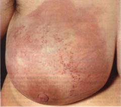
Diffuse seals are similar to the acute form of mastitis or mastopathy. They come in five varieties:
- edematous, more often during pregnancy and breastfeeding. The skin of the breast swells and becomes saturated with infiltrate, turns red and looks like a lemon peel. Edema appears due to compression of the infiltrate of the milk ducts;
- armored with characteristic tissue infiltration and spread to the chest. The skin becomes dense, bluish-red, inactive and wrinkled. In it, you can feel a lot of nodules, find ulcerations and a shell crust;
- erysipelatous (inflamed) with focal redness, swollen jagged edges. The skin of the chest wall is involved in the inflamed process. Accompanied by inflammation high temperature up to 40C and fever. It is badly treated.
- Vastitis-like with an increase in the area on the skin, tension, redness and an increase in local temperature in the compaction zone. They will be dense, slightly mobile and palpable under the fingers in all areas. Characterized by rapidly spreading inflammation accompanied by fever.
- in the form of psoriasis or eczema (with Paget's disease) accompanied by bright hyperemia, engorgement of the nipple and areola with the appearance of first dry, then weeping crusts and scabs on them, and under them - wet granulations. The spread of carcinogenesis will take place through the milk ducts deep into the breast.
Informative video on the topic: "3 main signs of breast cancer"
Metastases in breast cancer
Metastases of breast cancer appear when single tumor cells spread through the bloodstream (hematogenous way) and lymphatic fluid (lymphogenic ways) during the period early development oncogenic tumor. The rapid occurrence of secondary tumors due to metastases occurs only in the case of a depleted immune system of the body, especially in aggressive forms of cancer.
With high immunity, the body prevents the reproduction of cancer cells outside the mammary glands and metastatic foci are not formed. A tumor that does not go beyond the place of its formation: the mammary gland or duct is called non-invasive.
If the tumor enlarges with uncontrolled growth and spreads beyond the lobule or duct of the breast, it is called invasive (invading).
With the expression of tumor cells - ErbB-2 proteins, metastasis begins. Therefore, immunological analysis of breast biopsy can show this expression in order to confirm the aggressiveness of the early stage of the disease, before metastases appear. When metastases are detected by scintigraphy or PET-CT, it is already possible to indicate the spread of cells in the tissues of the liver, brain, lungs, and bones.
Breast cancer, metastases can be detected both in the initial stages of neoplasm development and after its recurrence. Tumor metastases often remain in a latent (dormant) state for a long time. After the removal of the primary tumor formation, they tend to "sleep" for 7-10 years and appear only under the influence of provocative factors.
The site of metastasis development is the nearest (regional) lymph nodes - anterior thoracic, axillary, subclavian, supraclavicular and parasternal. As the cancer progresses, the lymph nodes increase in size, which is called lymphadenopathy.

Regional lymph nodes are no longer able to prevent further metastasis of cancer cells, so hematogenous metastases reach:
- brain and spinal cord;
- liver and kidneys;
- lungs;
- spongy bones.
When cancer cells enter these organs, the tumor island increases to the size of a metastasis, and manifests itself with the following symptoms:
- in the brain- headache, general and muscle weakness in the limbs, visual impairment: double vision or loss of visual field, psychological disorders, decreased level of consciousness, convulsions;
- in the spinal cord- pain and numbness, paresthesia and muscle weakness, symptoms of a dangling hand and a slapping foot, Horner's syndrome may be observed in the brachial plexus;
- in the liver- heaviness and bloating of the abdomen, accompanied by prolonged pain, the development of jaundice with a decrease in liver tissue capable of functioning, weight loss;
- in the kidneys- blood in the urine, hematuria, fatigue, sudden weight loss, lack or decrease in appetite, high sweating, high fever, bouts of back pain, anemia, impaired hormone production and consequently a decrease in red blood cells, high blood pressure;
- in the lungs- persistent cough: dry and wet, shortness of breath during exertion and at rest;
- in spongy bones- steadily increasing pain in the back (vertebrae), pelvic bones and large joints, including knee and ankle, hip and shoulder. When the roots of the spinal nerves are compressed by the affected vertebrae (more often in the lumbar region), the symptoms are manifested by numbness or weakness of the limbs, a violation of the physiological activity of the intestines and bladder: fecal and urinary incontinence develops.
Stages of breast cancer and their classification
When determining five stages of breast cancer (from 0 to 4), a treatment regimen for patients is outlined and the effectiveness of recovery is predicted.

determined by the following factors:
- tumor size (T1, T2, T3, T4);
- invasiveness of education;
- damage to the lymph nodes (N 0, N1, N2, N3);
- the presence of metastases in other organs - M0, (absent) M1 (available).
Stages of breast cancer - classification:
| Stage | Size, see | The defeat of the lymph nodes | distantmetastases |
| 0 | Absent | Missing | |
| I | T1 = 2 | No metastases - N 0 | Not identified - M 0 |
| II | T2 = 3-5 | N 1 - metastases of I-II level were detected in the lymph nodes on the one hand, palpable | M 0 or M 1 - absent or there are single distant metastases |
| II-A | T2 = 2 or 2-5 | Affected lymph nodes under the armpits, lymph nodes are not affected |
«» «» |
| II-B | T3= 2-5 or T3>5 | Lymph nodes are affected., lymph nodes are not affected. | |
| III | T 3 >5 | N 2 metastases of I-II level were detected in the lymph nodes in the cavity under the armpits | M 0 or M 1 - absent or there are distant metastases. |
| III-A | Any | Under the armpits soldered lymph nodes | «» |
| III-B | Any | Grows into the skin of the breast, lymph nodes are soldered under the armpits | «» |
| III-C | Any | Lymph nodes below and above the collarbone are affected and/or the tumor has grown into the chest | «» |
| IV | Any | The tumor spread beyond the borders of the breast, there are nodules and ulcerations on the skin, N 3 - level III metastases on both sides of the chest, under the breast, under the armpits, above the collarbone, palpable | M 1 - there are multiple metastases in any organ and bones |
Early stages of breast cancer - 1, II-A, II-B and III-A.
After the operation, the treatment of stage 1 breast cancer lasts 2-3 weeks. To say about life expectancy, its degree is determined within 10 years after the end of therapy. If stage 1 breast cancer is diagnosed, the prognosis is positive, the 5-year survival rate exceeds 85% of all cases. If stage 2 breast cancer is identified, life expectancy over 5 years will account for about 66% of all cases.
Late stages of breast cancer - III-B, III-C and IV. The forecast is optimistic or negative. If stage 3 breast cancer is diagnosed, life expectancy of more than 5 years is 41% of all cases. This is possible in the presence of tumors over 5 cm with their germination in the tissues surrounding the breast, lesions of the lymph nodes under the arms and in other areas, but in the absence of metastases.
If the diagnosis is “stage 4 breast cancer”, patients will have a life expectancy of more than 5 years in only 10% of all cases. This is possible with a tumor size of more than 5 cm, the presence of lesions in the lymph nodes, and if metastases are detected in distant important organs.
Invasive breast cancer, ductal and lobular. The level of estrogen and progesterone in the body of the mammary gland, a specific protein HER2 / neu, indicates the type (form) of cancer.
The condition of women varies depending on the hormonal background. For them, the hormones that the ovaries produce are important. Natural physiological processes occur under the influence of estrogens, progesterone, pituitary hormones - LH, FSH.
Many forms of breast hyperplasia occur with endocrine disorders and high levels of estrogens and prolactin with a reduced level of progesterone. Breast cancer can manifest itself at the same ratio and be estrogen-dependent and progesterone-dependent.
For hormonal imbalance in the treatment, endocrine therapy is used. The effectiveness of treatment is 75%. At the same time, ovarian function is regulated and physical (irradiation) and surgical castration is used.
Negative cancer is the most severe form because it is difficult to treat. It is called triple-negative breast cancer because of the presence of receptors for one of three proteins in the body, such as estrogen, progesterone, and the specific tumor protein HER2/neu.
There are two types of estrogen-dependent cancer, A and B.
Luminal cancer type A women can get sick during menopause in 30-40% of all cases. Hormone cells: estrogen and progesterone will be well perceived by tumor cell receptors, but cells of the HER2/neu tumor protein will not be perceived at all. Their sensitivity to the breast cancer cell growth marker will be low - Ki67.
Luminal cancer is well treated with hormone therapy with Tamoxifen (an estrogen antagonist) and an aromatase inhibitor, an adrenal enzyme that promotes the conversion of testosterone to estrogen. At the same time, relapses are reduced, and the percentage of cure is increased.
Luminal cancer type B women of childbearing age get sick (14-18%). Cancer is characterized by frequent relapses accompanied by metastases to the lymph nodes. The disease is difficult to treat, it is poorly amenable to hormone and chemotherapy. In rare cases, immunotherapy (stimulation of immunity) with the help of the drug Transtuzumab, human monoclonal antibodies to the tumor protein HER2 / neu, stops cell growth.
Infiltrating cancer comes in several forms:
- two forms of non-invasive cancer in the ducts and lobules of the breast;
- two forms of invasive (infiltrating) cancer in the ducts and lobules;
- histological form of cancer: metaplastic, papillary, colloidal, medullary.
With infiltrating cancer, flows and lobules are affected, and in 70% they have symptoms of ductal carcinoma. The tumor may have the appearance of a dense potato-like formation.
If poorly differentiated cells are detected, then the course of the disease is characterized by aggressive symptoms, accompanied by metastases in the armpit and damage to the lymph nodes.
The most severe is the mixed form with histological changes in the lobules and ducts. Treatment is carried out surgical removal and chemotherapy.
Diagnosis of breast cancer in women
Breast cancer is diagnosed at an early stage. The doctor examines the patient in a standing position. At the same time, they release and raise their hands so that he can assess the contours, size, symmetry and condition of the skin of the chest. The doctor may identify:

- how much the nipple has shifted, deformed and its level has changed;
- the presence of pathological wrinkling of the skin of the nipple, swelling, hyperemia and discharge;
- when palpating the lymph nodes under the armpits, above and below the collarbones - the presence of a lesion (enlargement of the node);
- when palpating the gland - the consistency and structural homogeneity of the gland.
Diagnosis of breast cancer includes tests to rule out (or confirm) Hodgkin's disease, cancers in the lungs, ovaries, pancreas, and to identify skin conditions like squamous cell carcinoma. In some cases, a blind mastectomy is performed - the mammary gland is removed without a cytological examination.
After a clinical examination, the diagnosis is confirmed on the basis of indications:
- mammography (x-ray of the mammary glands);
- ultrasound examination (ultrasound) to determine the nature of the formation: solid or cystic;
- puncture biopsy - cytological examination of breast tissue;
- aspiration biopsy and subsequent cytological examination of the aspirate;
- selective excisional biopsy of formations located deep.
If there are estrogen and progesterone receptors in the biopsy, then hormone therapy is used to treat receptor-positive tumors. After it, the prognosis improves even with stage 3 breast cancer.
To determine diploidy (with DNA index = 1.00) or aneuploidy (with DNA index + 1.00) and the fraction of cells in the S-phase of mitosis, duct cytometry is performed. High fraction aneuploid tumors worsen the prognosis after treatment.
To determine metastases and if a recurrence is suspected, oncomarkers of breast cancer are used: CEA, CA15-3, CA 27-29 and their level is determined. Since it is necessary to examine a large area of the body when looking for metastases, scintigraphy of the skeletal system is performed with simultaneous examination of single suspicious nodes using X-rays.
A tumor marker for breast cancer is used to confirm the diagnosis along with classical research methods:
- Ultrasound of the abdominal organs;
- MRI of the brain and spinal cord;
- computed tomography of the brain, pelvis, abdomen, chest;
- PET-CT.
The article presents an informative video on the topic: “Breast Cancer: Risk Factors, Symptoms, Diagnosis, Treatment Options”
Methods of treatment of breast cancer
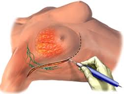
Surgical treatment of breast cancer is carried out taking into account the stage of the disease, the size and location of the tumor in the breast, the number of oncogenic neoplasms, the shape and size of the breast. The question of the availability of technical probabilities for radiation therapy and the operation itself, the possibility of preserving the mammary gland is considered.
Treatment of breast cancer with a modified radical mastectomy will save the mammary gland. Tylectomy is performed to correctly assess the extent of the tumor and improve the cosmetic result.
Contraindications to organ-preserving operations on the mammary gland are:
- large tumors on small mammary glands;
- primary tumors located near the nipple;
- multiple tumors in the breast;
- contraindication to radiation therapy;
- late treatment (after stage 2);
- microcalcifications in the duct or a large lesion within it.
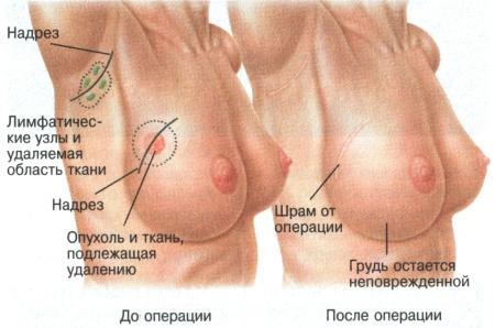
Held palliative or radical. In this case, in the case of multifocal cancer, the entire affected breast and lymph nodes under the arms are removed.
Lumpectomy (sectoral resection), Lymphadenectomy of the lymph nodes under the arms (1 and 2 levels), irradiation (after surgery) is performed when small primary tumors (less than 4 cm) and intraductal carcinoma are detected.
Also carried out:
- mastectomy:
- simple (Maden operation): the breast is removed near the nipple and lymph nodes of the 1st level;
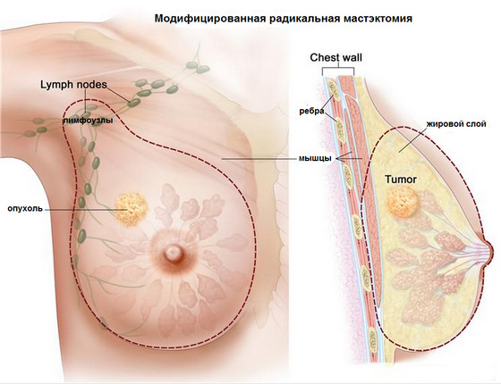
- modified radical (Patey operation): skin is removed within the breast, mammary gland, pectoralis minor muscle and fatty tissue, lymph nodes under the armpits, above and below the collarbone;
- radical Halsted operation: tissue is removed as in Patey's operation and the pectoralis major muscle, but the pectoral nerve is preserved to prevent denervation of the serratus anteriorly and eliminate the symptom of the pterygoid scapula;
- extensive and radical, during which mediastinal lymph nodes, large or medially located tumors with the presence of parasternal (inside the chest) metastases are removed;
- reconstructive surgery using subpectoral prosthetics.
Breast reconstruction is combined with a mastectomy or it is performed after the first surgical wound has healed.
When diagnosed with breast cancer, how long do they live after surgery? All patients want to know about this, but hardly anyone can give a definite answer. The prognosis depends on the age, location, degree of invasion and spread of the tumor, stage, histological parameters, operability (complete or partial removal of the tumor) and concomitant diseases. The most favorable prognosis will be with a completely removed primary lesion and regional lymph nodes, no metastases, a positive response after a course of chemotherapy and no recurrence within a year after surgery and treatment.
Conducting radiation therapy
There are three types of radiotherapy for breast cancer. Spend:
- external beam therapy;
- radiotherapy with modulated intensity;
- brachytherapy (internal or interstitial using a balloon or catheter). Used as an independent method of treatment or additional after surgery.
Here you can find out how it is done. The mammary gland and metastasis zones in the regions of the body are irradiated before the operation, after it - the mammary gland and lymph nodes, subject to the presence of metastases. Radiation therapy after surgery is carried out by those who have not been performed before it, as well as patients with risk factors:
- tumors (primary) more than 5 cm;
- metastases in 4 or more lymph nodes under the armpits;
- penetration of the tumor into the fascia and / or muscle of the chest, reaching the resection line, spreading into the fatty tissue under the armpits from the lymph nodes.
The classic effects of radiation therapy for breast cancer, such as hair loss and persistent nausea, are not present due to the very small dose of ionizing radiation. Acute radiation sickness will not develop.
Side effects in the middle of the course are manifested:
- general fatigue that lasts 1-2 months after therapy;
- episodic short-term attacks of pain in the gland: sharp shooting (rarely) and dull aching;
- radiation dermatitis: local irritation of the skin of the breast after 3-4 weeks, accompanied by swelling of the subcutaneous tissue, redness, itching, dry skin or dermatitis in the form sunburn, in which the epidermis exfoliates, and moist blisters form (more often under the breast and under the armpits).
The consequences of irradiation that do not require additional treatment are manifested:
- moderate swelling disappearing after 6-12 months;
- bronzing (darkening) of the skin;
- moderate pain discomfort in the chest and muscles around them due to myositis after irradiation.
Important! Require treatment for complications that manifest themselves:
- lymphedema (edema) of the upper limb after irradiation of the lymph nodes under the armpits and lymphadenectomy (surgery to remove the lymph nodes)
- severe paresthesia with chronic pain syndrome against the background of loss of muscle strength of the upper limb, including the hand, due to degeneration of nerve fibers;
- radiation pneumonitis - reactive inflammation of the lungs after x-ray exposure (after 3-9 months);
- radiation ulcers on the skin of the breast. They may need surgical treatment.
Conducting chemotherapy
Adjuvant with an increased risk of distant metastasis is carried out in conjunction with radiation therapy to slow or prevent recurrence, improve survival in patients with or without lymph node metastases.
Combined chemotherapy for breast cancer is more often performed than monotherapy, especially with metastases. Conduct six monthly courses. Treatment is performed with drugs tested for toxicity.
Maximum doses are prescribed, for example:
- three drugs at once: Fluoruracillus, Methotrexate and Cyclophosphamide (Cyclophosphamide);
- with frequent relapses or metastases - Fluorouracill, Doxorubicin hydrochloride and Cyclophosphamide;
- with metastases - Taxol (Paclitaxel), Vinblastine, Thiophosphamide, Doxorubicin.
Do not carry out radiation therapy for the reason:
- pregnancy;
- previously received exposure to another organ;
- connective tissue diseases: lupus erythematosus, systemic vasculitis, scleroderma, against which the patient will suffer from increased sensitivity to procedures;
- the presence of concomitant diseases: severe diabetes mellitus, cardiovascular insufficiency, anemia.
The classic side effects of chemotherapy for breast cancer are:
- lack of appetite due to nausea and vomiting;
- indigestion, diarrhea and constipation;
- apathy, weakness, lethargy and loss of strength;
- hair loss (alopecia);
- fever and fever;
- a decrease in the body's defenses and the activation of chronic diseases, the emergence of acute new diseases;
- inhibition of the functional work of the ovaries;
- anemia and decreased hemoglobin levels;
- leukopenia (decrease in the number of leukocytes) and thrombocytopenia (decrease in the number of platelets) in the blood.
Carrying out hormone therapy
Adjuvant hormone therapy for breast cancer is prescribed if:
- a long period (more than 5 years) without the formation of metastases;
- elderly patients;
- the presence of metastases in bone tissues;
- development of minimal metastases in the lungs and multiple regional ones;
- histological confirmation of I and II degrees of cancer;
- a long period of remission after hormone therapy performed earlier.
Hormone therapy for breast cancer is effective after chemotherapy and in the case of progesterone receptors (PR +) and estrogen receptors (ER +) on cancer cells.
Treatment of patients during premenopause is carried out with drugs, such as:
- Tamoxifen, Luliberin antagonists: Leuprolide acetate, Aminoglutethimide, Hydrocortisone.
Treatment of patients in the postmenopausal period is carried out with drugs, such as:
- Tamoxifen, Megestrol acetate, Aminoglutethimide;
- high doses of estrogen - Diethylstilbestrol, Luliberin antagonists.
In the presence of ERc-positive tumors, it is preferable to treat with Tamoxifen. In ERc-negative tumors, Tamoxifen is less effective. Also, treatment is carried out with aromatase enzyme inhibitors, Zoladex (Goserelin) and oophorectomy (removal and/or irradiation of the ovaries). After an oophorectomy, a woman becomes infertile. Side effects are manifested by redness and dryness of the skin, dryness in the vagina, a sharp change in mood.
Conducting targeted therapy
Targeted therapy for breast cancer is one of the new developments in cancer treatment. Its difference from the above types of treatment is the absence of side effects on body tissues and the rapid destruction of the tumor. The treatment is carried out with targeted drugs (point exposure) that affect the molecule that promotes the growth of tumor cells. This treatment is called "molecular targeted therapy" because it blocks the growth of tumor cells and starts the process of their destruction. It is often combined with chemotherapy and radiotherapy.
Before using targeted therapy, tests are performed to determine receptor sensitivity by immunohistopathological examination of tumor tissue removed during a biopsy or during surgery.
Immunohistochemistry is used to determine the number of HER-2 receptors, estrogen and progesterone on the surface of tumor cells.
Therefore, the treatment is carried out with the following drugs:
- Tamoxifen, Toremifene (Fareston), Fulvestrant (Fazlodex);
- drugs that affect ER-positive tumors, such as: Anastroizol (Arimidex), Letrozole (Femara), Exemestane (Aromasin) - inhibitors of the aromatase enzyme that produces estrogens;
- selective growth factor blockers: Bevacizumab (Avastin), Panitumumab (Vectibix), Cetuximab (Erbitux), Trastuzumab (Herceptin). They block angiogenesis (vascular growth) and inhibit the development of a network of vessels around tumor cells, thereby slowing down tumor growth.
Damaged DNA in cells is repaired with inhibitors (blockers) of the PARP protein, after which the apoptosis program (“cell death”) is activated with drugs: Veliparib, Iniparib, Olaparib, provided that there are no such main receptors in the cells as:
- Her-2 (epidermal growth factor);
- estrogen receptor ER;
- progesterone receptor PR.
The prognosis of targeted therapy for breast cancer is optimistic. It is used as a prevention of possible recurrence and to control the spread of metastases. The use of drugs allows patients to live long with cancer without compromising the quality of life.
Carrying out immunotherapy
With immunotherapy, cancer cells can be marked and made visible to immune cells. It can directly kill reborn cells or strengthen the immune system.
Immunotherapy of breast cancer is performed by non-specific vaccination: the use of BCG, stimulation of phagocytic activity with the help of a protein derivative of tuberculin, the inclusion of Timidrin in leukocytes, etc.
It is important to know! Immunotherapy:
- restores and normalizes immune-protective mechanisms, if reduced indicators of immunity are found: humoral and cellular;
- used after surgery, radiation and chemotherapy, if as a result of this stress has occurred and the body's reactivity has been impaired;
- used for distant metastases: manifesting and subclinical to prevent the appearance of a secondary tumor.
Treatment with drugs showed itself well: Levimezol, Zimozan, Prodigiozan. At the same time, specific and nonspecific immunity factors were activated. Restored immunity contributes to a long relapse-free period after mastectomy.
In the case of relapses and metastases, immunotherapy helps to increase the frequency of regression of cancer foci. With persistent inhibition of immunoreactivity in patients, immunotherapy will not bring high results.
Disease prevention
Prevention of breast cancer includes self-examination of the breast after menstruation. Should:
- timely conduct conservative therapy of fibrocystic mastopathy;
- to be observed annually by a gynecologist-mammologist, especially after 30-40 years;
- women 40-50 years old to undergo mammography annually or once every 2 years;
- women 50 years of age with risk factors - annually examine the breast with a mammogram;
- wear a comfortable bra with wide straps so that there are no chafing and redness, especially during menstruation with swelling of the chest;
- lead a healthy lifestyle, including a healthy diet;
- protect the chest from direct sunlight, injuries and surgical interventions.
Informative video: modern view of breast cancer
Be healthy!
Oncopathology of the breast is an ailment that occurs as a result of the degeneration (mutation) of mammary gland cells. Breast cancer cannot be called a purely female pathology. Although it is not difficult to detect the first signs of cancer on your own, the tumor is most often detected during examination by a gynecologist. The earlier deviations are detected, the less traumatic treatment methods will be applied.
Causes
The development of cancer is due to the mutation of certain genes (DNA) that are responsible for inhibiting tumor growth. Women at risk for breast cancer include:
- having a burdened heredity (the presence of cancer in previous generations increases the risk up to 25%);
- nulliparous, age primiparous (the first birth occurred after 30 years);
- older than 50 years (during menopause);
- having a history of abortion;
- refused breastfeeding;
- long-term use of hormonal contraceptives;
- who have undergone mastopathy (degenerated from a diffuse form into a nodular one), mastitis;
- suffered injuries, bruises, hypothermia of the mammary glands;
- with marked early puberty;
- with endocrine pathology (including obesity and diabetes);
- previously received courses of radiation therapy;
- smokers, alcohol abusers;
- living in areas with high background radiation;
- often experiencing stress.
Breast cancer in men is extremely rare (male incidence is only 1% of female), most often provoked by hormonal imbalance (gynecomastia - growth of the mammary glands due to an increase in the amount of female hormones in the male body).
Symptoms
Women usually do not attach importance to mild soreness and discomfort in the chest, however, it is these signs that may indicate the development of a tumor. A breast with cancer looks like this (one or more external signs):
- the area of the skin has become hard, the skin is in the form of a "lemon peel";
- local redness, swelling;
- the appearance of retraction on the skin;
- deformation of the mammary gland (enlargement, asymmetric "ripple");
- the appearance of a venous pattern on the skin of the mammary glands;
- nipple retraction;
- pathological discharge from the nipple (serous, may be bloody);
- the appearance of an eroded area on the chest;
- detection of compaction on palpation.
Important! If you experience the above symptoms and pain visiting a mammologist is a must.
Long-term symptoms of breast cancer:
- a palpable solid tumor, sometimes a pasty nodule;
- the appearance of nodes in the second gland (the risk of spreading to the second breast increases 4 times);
- an increase in axillary, supraclavicular and subclavian lymph nodes (stage 3 tumor);
- chest pain (only in 15% of cases of breast cancer);
- general symptoms (weakness, subfebrile condition, etc.);
- distant metastases (stage 4 of the oncological process).
Stages of breast cancer:
- 0 stage. Non-invasive tumor, cancer cells are limited to the focus.
- 1 stage. Altered cells grow into neighboring tissues, damaging the glandular. Tumor up to 2 cm in diameter.
- 2 stage. Compaction up to 5 cm, the lymphatic system is included in the oncoprocess.
- 3a stage. Node more than 5 cm, extensive damage to the lymph nodes.
- 3b stage. The size of the main node does not matter, the oncoprocess spreads to the chest, affecting the internal lymph nodes.
- 4 stage. It is characterized by distant metastases in the lungs, liver, neck organs.
Important! Detection of cancer at stage 0-2 guarantees a 5-year survival rate of 70%. Treatment in Germany and other foreign countries gives higher incidence rates - from 90 to 100%.
Self-examination of the mammary glands

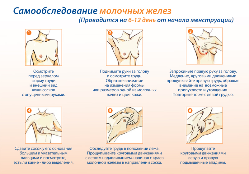
Every woman should know the rules of breast examination and conduct regular self-exams in order to detect breast cancer in a timely manner.
Looking at the mirror
A woman in front of a mirror examines her breasts for pathological changes in the size, shape of the breasts, skin and nipples, in the armpits. The examination is carried out in two positions: with the arm thrown back behind the head from the side of the examined organ, with the arm extended along the body. Pressing around the gland and squeezing the nipple should be gentle. The examination of the glands can be repeated in the shower. Sliding the fingertips over wet skin, the mammary gland is carefully examined in circular motions.
Important! The left mammary gland in women is physiologically somewhat larger than the right.
Lying examination
A pillow is placed under the shoulder blade on the side being examined. With three middle fingers, carefully examine the chest in a circle from the periphery to the center, starting from the sternum. The duration of the examination is about 5 minutes.
Diagnostics
Diagnosis of oncological diseases of the mammary glands includes the following examinations:
- medical examination of the breast by a gynecologist / mammologist;
- Ultrasound examination of the mammary glands (will distinguish between cystic pathology and cancer);
- blood tests (oncomarkers - cancer is sometimes accompanied by their absence, immunological tests);
- mammography with the possible taking of tumor material for a biopsy;
- the actual biopsy is stereotaxic / vacuum (determines the type of tumor cells).
To recognize cancer that has metastasized to other systems and organs help:
- X-ray of the chest, bones;
- computed tomography (scans the damaged organ in layers);
- positron emission tomography of organs with suspected metastases and lymph nodes.
Treatment
The treatment regimen is determined by a mammologist-oncologist according to the stage of the oncological process, general condition and sensitivity to hormone therapy.
Treatment strategy:
Radiation therapy
Cyberknife - modern and enough effective method radiosurgical removal of the tumor by exposure to a powerful gamma beam directly on the tumor. Radiation therapy courses can precede or supplement surgical intervention at any stage of the oncological process.
Chemotherapy
The most popular cytostatic is Tamoxifen. Aromasin courses are less commonly prescribed due to its strong side effects.
hormone therapy
Up to 75% of breast cancer is represented by hormone-dependent tumors. Treatment with individually selected doses of estrogens or progesterone is carried out before surgery on tumors large sizes, with a preventive purpose to prevent relapses and for therapeutic purposes, even with inoperable patients.
Targeted Therapy
The essence of the technique lies in the point impact: the drugs are delivered directly to the tumor, do not affect healthy cells.
Immunotherapy
Taking specific anticancer drugs (Herceptin) allows you to stimulate the immune system to fight altered cells.
Operation
More often, partial removal of the breast (the lobe in which the tumor has developed) is acceptable. Complete removal of the breast (mastectomy) is indicated when the entire gland is involved in the oncological process. To eliminate psychological discomfort, women are offered subsequent prosthetics with implants. In stage 4, surgery is often inappropriate.
The maximum healing effect is achieved with combined treatment and individual selection of dosages.
In the United States, a healthy woman who has breast cancer among her relatives and who has given birth to at least two children may own will decide on a prophylactic mastectomy followed by prosthetics.
Breast cancer is not a death sentence. According to Western experts, 70% of women do not require radical removal of the breast, subject to early detection of cancer. An annual medical examination, self-examination of the breast and an immediate visit to the clinic allows diagnosing the tumor process at an early stage, which requires less radical treatment and significantly increases the survival rate. However, if pathological signs are found, you should not sound the alarm, often the seals found are mastopathy or a benign node.
Breast cancer (carcinoma)- the most common malignant tumor of the mammary glands.
The disease is characterized by a high prevalence. In developed countries, it occurs in 10% of women. Europe is leading the way. Japan has the lowest prevalence of breast cancer.
Some epidemiological data on breast cancer:
- most cases of the disease are registered after the age of 45 years;
- after 65 years, the risk of developing breast carcinoma increases by 5.8 times, and compared with a young age (up to 30 years) it increases by 150 times;
- most often the lesion is localized in the upper outer part of the mammary gland, closer to the armpit;
- 99% of all patients with breast carcinoma are women, 1% are men;
- isolated cases of the disease in children are described;
- mortality in this neoplasm is 19 - 25% of all other malignant tumors;
- Breast cancer is one of the most common tumors in women today.
At the moment, there is an increase in the incidence worldwide. At the same time, in a number of developed countries there are downward trends due to well-organized screening (mass examination of women) and early detection.
Causes of breast cancer
Exists a large number of factors contributing to the development of breast carcinoma. But almost all of them are associated with two types of disorders: increased activity of female sex hormones (estrogens) or genetic disorders.Factors that increase the risk of developing breast cancer:
- female;
- unfavorable heredity (the presence of cases of the disease in close relatives);
- beginning earlier than 12 years or ending later than 55 years, their presence for more than 40 years (this indicates increased estrogen activity);
- absence or its onset for the first time after 35 years;
- malignant tumors in other organs (in the uterus, ovaries, salivary glands);
- various mutations in genes;
- the effect of ionizing radiation (radiation): radiation therapy for various diseases, living in an area with an increased radiation background, frequent fluorography for tuberculosis, occupational hazards, etc .;
- other diseases of the mammary glands: benign tumors, nodular forms of mastopathy;
- the action of carcinogens (chemicals that can provoke malignant tumors), some viruses (so far, these points have been poorly studied);
- tall woman;
- low physical activity;
- abuse , ;
- hormone therapy in high doses and for a long time;
- continuous use for contraception;
- after .
Usually, malignant tumors of the mammary glands are heterogeneous. They consist of different types cells that multiply at different rates respond differently to treatment. In this regard, it is often difficult to predict how the disease will develop. Sometimes all symptoms grow rapidly, and sometimes the tumor grows slowly, without leading to noticeable disturbances for a long time.
The first signs of breast cancer
 Like other malignant tumors, breast cancer is very difficult to detect at an early stage. For a long time, the disease is not accompanied by any symptoms. Its symptoms are often discovered by accident.
 Symptoms that require immediate medical attention:
- breast pain that has no apparent cause and persists for a long time;
- feeling of discomfort for a long time;
- seals in the mammary gland;
- change in the shape and size of the breast, swelling, deformation, the appearance of asymmetry;
- nipple deformities: most often it becomes retracted;
- discharge from the nipple: bloody or yellow;
- changes in the skin in a certain place: it becomes retracted, begins to peel off or wrinkle, its color changes;
- a dimple, a depression that appears on the mammary gland, if you raise your hand up;
- swollen lymph nodes in the armpit, above or below the collarbone;
- swelling in the shoulder, in the region of the mammary gland.
- Regular self-examination. A woman should be able to properly examine her breasts and identify the first signs of a malignant neoplasm.
- Regular visits to the doctor. It is necessary to visit a mammologist (specialist in breast diseases) at least once a year.
- Women over the age of 40 are recommended to undergo regular X-ray examination aimed at early detection of breast cancer.
How to examine your breasts on your own?
Self-examination of the mammary glands takes about 30 minutes. It should be done 1-2 times a month. Sometimes pathological changes are not immediately felt, so it is advisable to keep a diary and note in it the data, your feelings based on the results of each self-examination.Inspection of the mammary glands should be carried out on the 5th - 7th day of the menstrual cycle, preferably on the same days.
visual inspection
 This should be done in a warm, bright room with a mirror. Undress to the waist and stand exactly in front of the mirror, so that you can clearly see the howling chest. Relax and even out your breathing. Pay attention to the following points:
 - Are the right and left mammary glands symmetrical?
- has one mammary gland increased compared to the other (it is worth remembering that normally the sizes of the right and left mammary glands may differ slightly)?
- does the skin look normal, are there any suspicious areas with a changed appearance?
- do the nipples look ok?
- nothing else suspicious seen?
Feeling
 Feeling the chest can be carried out in a standing or lying position, whichever is more convenient. If possible, it is better to do this in two positions. The examination is carried out with the fingertips. The pressure on the chest should not be too strong: it should be sufficient so that changes in the consistency of the mammary glands can be felt.
 First, one mammary gland is felt, then the second. Start from the nipple, then move the fingers outward. For convenience, you can feel in front of a mirror, conditionally dividing the mammary gland into 4 parts.
Moments to pay attention to:
The general consistency of the mammary glands - has it become denser since the last examination?
- the presence of seals, nodes in the tissue of the gland;
- the presence of changes, seals in the nipple;

If changes are found, contact one of the specialists:
 With the help of self-examination, it is possible to detect not only breast cancer, but also benign neoplasms, mastopathy. If you find something suspicious, then this does not indicate the presence of a malignant tumor. An accurate diagnosis can only be established after examination.
 For the purpose of early diagnosis of breast cancer, women over 40 are recommended to undergo three studies annually:
- Mammography - X-rays of the breast. Existing seals in the tissue are revealed. The modern method is digital mammography.
- Determination of the level of female sex hormones - estrogens. If it is high, there is an increased risk of developing breast cancer.
- Oncomarker CA 15-3 is a substance that is produced by breast carcinoma cells.
Symptoms and appearance of different forms of breast cancer
| Nodular form of breast cancer | A painless dense formation is palpated in the thickness of the mammary gland. It can be round or have an irregular shape, it grows evenly in different directions. The tumor is soldered to the surrounding tissues, therefore, when a woman raises her hands, a depression forms on the mammary gland in the corresponding place. The skin in the area of the tumor is wrinkled. In the later stages, its surface begins to resemble a lemon peel, ulcers appear on it. Over time, the tumor leads to an increase in the size of the breast. What does nodular breast cancer look like? |
| Edema-infiltrative form | This form of breast cancer is most common in young women. Pain is often absent or mild. There is a seal that occupies almost the entire volume of the breast. Symptoms:
 |
| shell cancer | The tumor grows through the entire glandular tissue and adipose tissue. Sometimes the process goes to the opposite side, to the second mammary gland. Symptoms:
|
| Paget's cancer | A special form of breast cancer occurs in 3-5% of cases. Symptoms:
|
Breast cancer grades
The degrees of breast cancer are determined according to the generally accepted TNM system, in which each letter has a designation:- T is the state of the primary tumor;
- M - metastases to other organs;
- N - metastases in regional lymph nodes.
| The degree of the tumor process | Main characteristics |
| T x | The doctor does not have enough data to assess the condition of the tumor. |
| T0 | No tumor was found in the breast. |
| T1 | Tumor less than 2 cm in greatest dimension. |
| T2 | Tumor measuring 2 to 5 cm in greatest dimension |
| T3 | Tumor larger than 5 cm. |
| T4 | A tumor that has grown into the chest wall or skin. |
| N |
|
| N x | The doctor does not have enough information to assess the condition of the lymph nodes. |
| N0 | There are no signs indicating the spread of the process to the lymph nodes. |
| N 1 | Metastases in axillary lymph nodes, in one or more. In this case, the lymph nodes are not soldered to the skin, they are easily displaced. |
| N 2 | Metastases in the axillary lymph nodes. In this case, the nodes are soldered to each other or to the surrounding tissues, they are difficult to move. |
| N 3 | Metastases in peristernal lymph nodes on the affected side. |
| M |
|
| Mx | The doctor has no data that would help to judge tumor metastases in other organs. |
| M0 | There are no signs of metastases in other organs. |
| M1 | The presence of distant metastases. |
Of course, only a doctor after an examination can attribute a tumor to one or another stage according to the TNM classification. From this will depend on further tactics of treatment.
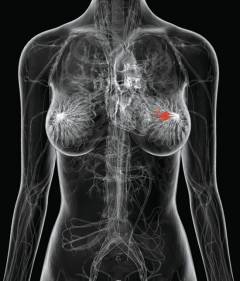
Classification depending on the location of the tumor:
- breast skin;
- nipple and areola (skin around the nipple);
- upper inner quadrant of the breast;
- lower inner quadrant of the breast;
- upper outer quadrant of the breast;
- lower outer quadrant of the breast;
- posterior axillary part of the mammary gland;
- the location of the tumor cannot be determined.
Breast Cancer Diagnosis
Inspection
 Diagnosis of malignant tumors of the breast begins with an examination by an oncologist or mammologist.
 During the examination, the doctor:
- ask the woman in detail, try to get the most complete information about the course of the disease, the factors that could contribute to its occurrence;
- will examine and palpate (palpate) the mammary glands in the prone position, standing with arms raised and lowered.
Instrumental diagnostic methods
| Diagnostic method | Description | How is it carried out? | |
| Mammography- a section of diagnostics that deals with non-invasive(without incisions and punctures) by examining the internal structure of the mammary gland. | |||
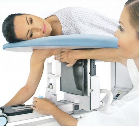
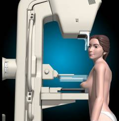
| X-ray mammography | the study of the mammary gland is carried out using devices that generate low-intensity radiation. Today, mammography is considered the main method of early diagnosis of malignant neoplasms of the breast. Has an accuracy of 92%. In Europe, X-ray mammography is mandatory for all women over the age of 45 on a regular basis. In Russia, it is mandatory for women over 40 years old, but in practice it is not carried out by all. With the help of X-ray mammography, tumors with a size of 2-5 cm are best detected. An indirect sign of a malignant neoplasm is a large number of calcifications - accumulations of calcium salts, which are well contrasted in the pictures. If they are found more than 15 per cm 2, then this is a reason for further examination. 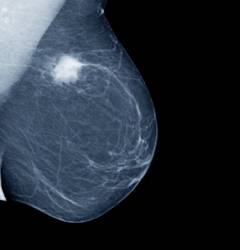 | The study is carried out in the same way as a conventional x-ray. The woman is stripped to the waist, leans against a special table, puts her mammary gland on it, after which a picture is taken. X-ray mammography devices must comply with the requirements established by WHO. Types of x-ray mammography:
 |
|
| MRI mammography | MRI mammography is a study of the mammary glands using magnetic resonance imaging. Advantages of MRI mammography over X-ray tomography:
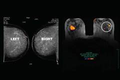 | Before the study, you must remove all metal objects from yourself. You can not take any electronics, as the magnetic field that the device generates can disable it. If the patient has any metal implants (pacemaker, joint prostheses, etc.), you need to warn the doctor - this is a contraindication to the study. The patient is placed in the apparatus in a horizontal position. She must be in a stationary position during the entire study. The time is determined by the doctor. |
|
| -mammography | Ultrasound examination is currently an additional method for diagnosing malignant neoplasms of the mammary glands, although it has a number of advantages over radiography. For example, it allows you to take pictures in different projections, does not have a harmful effect on the body. The main indications for the use of ultrasound diagnostics in breast cancer:
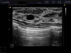 | The procedure is no different from conventional ultrasound. The doctor uses a special sensor that is applied to the mammary gland. The image is transmitted to the monitor, can be recorded or printed. Dopplerography and duplex scanning may be performed during an ultrasound examination of the mammary glands. |
|
| Computed tomomammography | The study is a computed tomography of the mammary glands. Advantages of computed tomomammography over x-ray mammography:
The study is worse than x-ray mammography, reveals small structures and calcifications. 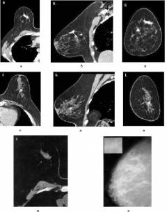 | The study is carried out in the same way as a conventional computed tomography. The patient is placed on a special table inside the apparatus. It must remain motionless throughout the study.  |
|
| Biopsy- excision of a fragment of breast tissue with subsequent examination under a microscope. |
|||
| Needle biopsy | The accuracy of the technique is 80 - 85%. In 20 - 25% of cases, a false result is obtained. | A fragment of breast tissue for research is obtained using a syringe or a special suction gun. The procedure is performed under local anesthesia. Depending on the thickness of the needle, there are two types of puncture biopsy:
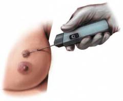 |
|
| Trepanobiopsy | Trepanobiopsy of the mammary glands is performed in cases where it is necessary to obtain more material for research. The doctor receives a piece of breast tissue in the form of a column. | Trepanobiopsy is performed using a special tool consisting of a cannula with a mandrel, into which a rod with a cutter is inserted. The intervention is performed under local anesthesia. The surgeon makes an incision in the skin and inserts a trepanobiopsy instrument through it. When the tip of the incisor reaches the tumor, it is pulled out of the cannula. With the help of a cannula, a column of tissues is cut off, and it is removed. After receiving the material, the wound is carefully coagulated to prevent the spread of cancer cells. During a study in the laboratory, it is possible to determine the sensitivity of tumor cells to steroid hormones (which include estrogens). This helps in the further choice of treatment tactics. 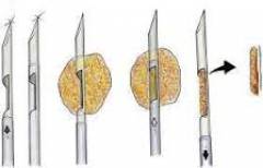 |
|
| Excisional biopsy | Excision - complete removal of the tumor with surrounding tissues. The entire mass is sent to the laboratory for analysis. This makes it possible to detect tumor cells at the border of the incision, to study the sensitivity of the tumor to sex hormones. | The surgeon removes the tumor with surrounding tissues during the operation. Thus, excisional biopsy is both a therapeutic and a diagnostic procedure. |
|
| Stereotactic biopsy | During a stereotactic biopsy, samples are taken from several different locations through a single needle. | The procedure resembles a conventional needle biopsy. It is always carried out under the control of x-ray mammography. The needle is inserted in a certain place, a sample is obtained, then it is sipped, the angle of inclination is changed and it is inserted again, this time in a different place. Multiple samples are obtained, making diagnosis more accurate. |
|

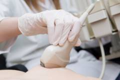
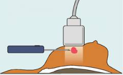
Laboratory methods for diagnosing breast cancer
| Study | Description | Methodology |
| Determination of the oncomarker CA 15-3 in the blood (syn.: carbohydrate antigen 15-3, carbohydrate Antigen 15-3, cancer Antigen 15-3) | Tumor markers are various substances that are determined in the blood during malignant neoplasms. Different tumors have their own tumor markers. CA 15-3 is an antigen located on the surface of the mammary ducts and secreting cells. Its content in the blood is increased in 10% of women with early stages of breast cancer and in 70% of women with tumors accompanied by metastases. Indications for the study:
| For research, blood is taken from a vein. Do not smoke for half an hour before taking the test. |
| Cytological examination of discharge from the nipple | If a woman has discharge from the nipple, then they can be sent for a laboratory test. When examined under a microscope, tumor cells can be detected. You can also make an imprint of the crusts that form on the nipple
| When studying secretions from the nipple under a microscope, cells characteristic of a malignant tumor are detected. |

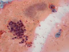
Breast Cancer Treatment*
Breast Cancer Treatment Methods:- surgical;
- chemotherapy;
- hormone therapy;
- immunotherapy;
- radiation therapy.
Surgery
 Surgery is the main treatment for breast cancer. Currently, oncological surgeons are trying to perform less voluminous interventions, to preserve breast tissue as much as possible, supplementing surgical methods with radiation and drug therapy.
 Types of surgical interventions for breast cancer:
- Radical mastectomy: complete removal of the mammary gland along with fatty tissue and nearby lymph nodes. This version of the operation is the most radical.
- Radical resection: removal of the breast sector along with subcutaneous fatty tissue and lymph nodes. Currently, surgeons increasingly prefer this variant of surgical intervention, since radical mastectomy practically does not prolong the life of patients compared to resection. The intervention must be supplemented with radiation therapy and chemotherapy.
- Quadrantectomy- removal of the tumor itself and surrounding tissues within a radius of 2 - 3 cm, as well as nearby lymph nodes. This surgical intervention can be performed only in the early stages of the tumor. The excised tumor must be sent for a biopsy.
- Lumpectomy- the smallest operation in terms of volume, during which the tumor and lymph nodes are removed separately. The surgical study was developed during the studies of the National Breast Surgery Augmentation Project (NSABBP, USA). The conditions for the intervention are the same as for quadrantectomy.
Radiation therapy
Types of radiation therapy depending on the timing:| Name | Description |
| Preoperative | Intensive short-term courses of irradiation are carried out. Goals of preoperative radiotherapy for breast cancer:
|
| Postoperative | The main goal of radiation therapy in the postoperative period is to prevent tumor recurrence. Places that are irradiated during postoperative radiotherapy:
|
| intraoperative | Radiation therapy can be used right during the operation if the surgeon tries to preserve the breast tissue as much as possible. This is useful at the stage of the tumor:
|
| Independent | Indications for the use of gamma therapy without surgery:
|
| Interstitial | The radiation source is brought directly to the tumor. Interstitial radiation therapy is used in combination with remote (when the source is at a distance) mainly in nodular forms of cancer. Purpose of the method: deliver as large a dose of radiation as possible to the tumor in order to destroy it as much as possible. |
Areas that may be exposed to radiation:
- directly the tumor itself;
- lymph nodes located in the armpit;
- lymph nodes located above and below the collarbone;
- lymph nodes located in the sternum.
Chemotherapy
 Chemotherapy- drug treatment of breast cancer, which uses cytostatics. These medicines destroy cancer cells and inhibit their reproduction.
 Cytostatics are drugs that have numerous side effects. Therefore, they are always prescribed strictly in accordance with established regulations and taking into account the characteristics of the disease.
The main cytostatics used in malignant tumors of the mammary glands:
- adriblastin;
- methotrexate;
- 5-fluorouracil;
- paclitaxel;
- cyclophosphamide;
- docetaxel;
- xeloda.
- CMF (Cyclophosphamide, Fluorouracil, Methotrexate);
- CAF (Cyclophosphamide, Fluorouracil, Adriablastine);
- FAC (Fluorouracil, Cyclophosphamide, Adriablastine).
hormone therapy
The main goal of hormone therapy is to eliminate the influence of female sex hormones (estrogens) on the tumor. Methods are used only in the case of tumors that are sensitive to hormones.Methods of hormone therapy:
| Method | Description |
| Removal of the ovaries | After the removal of the ovaries in the body, the level of estrogen drops sharply. The method is effective in a third of patients. It is used at the age of 15 - 55 years. |
"Medicinal castration" drugs:
| Medications suppress the release of follicle-stimulating hormone (FSH) by the pituitary gland, which activates the production of estrogen by the ovaries. The method is effective in a third of women aged 32 to 45 years. |
Antiestrogenic drugs:
| Antiestrogens are drugs that suppress the function of estrogens. Effective in 30% - 60% of women aged 16 to 45 years. |
Drugs that inhibit the aromatase enzyme:
| The aromatase enzyme is involved in the formation of steroid hormones, including the female sex hormones estrone and estradiol. By inhibiting aromatase activity, these drugs reduce estrogenic effects. |
Progestins (gestagens):
| Progestins are a group of female sex hormones that interact not only with their own receptors on the cell surface, but also with receptors designed for estrogens, thereby partially blocking their action. Drugs containing progestins, prescribed at the age of 9 to 67 years, have an effectiveness of 30%. |
| Androgens are preparations of male sex hormones. | Androgens inhibit the production of follicle-stimulating hormone (FSH), which activates the production of estrogen in the ovaries. The method is effective in 20% of girls and women aged 10 to 38 years. |
How does a doctor choose tactics for breast cancer treatment?
 The treatment plan for breast cancer is made individually.
 Features that the doctor should consider:
- the size of the neoplasm;
- the presence of metastases in the lymph nodes;
- germination in neighboring organs, the presence of distant metastases;
- laboratory data characterizing the cellular composition, the degree of malignancy of the tumor.
What alternative methods of treatment can be used for breast cancer?
Modern methods of treatment provide good prognosis in most women with malignant tumors of the mammary glands. So, at the beginning of treatment at stage I, about 95% of patients live longer than 5 years. Many have made a full recovery.Alternative methods are not able to provide an effective fight against the tumor process. Self-medication delays the visit to the doctor. Often such patients turn to a specialist when there are already distant metastases in the lymph nodes. At the same time, 70% of patients do not survive for 3 years.
The only right decision for a patient with suspected breast cancer is to see a doctor as early as possible, carry out diagnostics and, if necessary, start treatment in an oncology clinic.