The mucous membranes in the oral cavity are subject to skin diseases, and often become the area of primary manifestations of dermatological pathologies.
Dentistry involves the study of such phenomena, and sometimes the dentist is able to diagnose a dermatological diagnosis even before the symptoms of the disease appear on the skin.
One such problem is clear blisters that appear in the mouth or. This phenomenon is a real reason to worry about your health and a mandatory reason to visit a doctor.
The manifestations of such symptoms can be very diverse, and even identical symptoms in an adult and a child are not always provoked by the same infections or viruses. It is important to understand that you cannot postpone treatment “for later”!
The peculiarity of the formed blisters on the mucous membrane of the mouth is the fragility of their existence.
Having appeared, they soon open, forming erosion - the mucous membrane, unprotected by the top layer, becomes easily accessible to the influence (infection) of various microorganisms that are constantly present in the oral cavity.
The first signs are formed and become aggravated painful sensations, depriving the patient of peace, sleep, appetite and the very opportunity to eat food.
Manifestations of blistering dermatoses
According to Wikipedia, a “bubble” is considered to be a cavity element, up to 5 mm in diameter, resulting from a limited 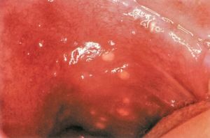 concentration inside any liquid.
A disease that combines clinical manifestations characterized by the formation of blisters on mucous membranes or non-inflamed skin is called pemphigus. Without adequate treatment, the pathology can cover large areas of the skin and has a malignant course.
The term “pemphigus” is applicable to a number of diseases of the mucous membranes, united by similar blistering rashes, but having different indicators (presence/absence) in smears of acantholytic cells, including clinical and pathological anatomical characteristics.
The initial symptoms are often localized in the mouth, in the absence of typical signs on the skin, which complicates diagnosis and can lead to misdiagnosis.
The classification of pemphigus in the oral cavity is divided into the following types:
True (acantholytic):
- vulgar;
- leaf-shaped;
- vegetative;
- seborrheic (erythematous), Senir-Usher syndrome.
False (non-acantholytic):
- bullous mucosineachial atrophying dermatitis (pemphigus of the eyes);
- Lever's bullous pemphigoid (nonacantholytic);
- benign non-acantholytic.
Causes and risk factors
Viral infections on the mucous membranes of the mouth are an extremely unpleasant and painful phenomenon. Most often, the damaging factor is viral, a common infection that combines a whole list of diseases provoked by various viruses:
- simple ;
- chicken pox;
- flu;
- parainfluenza;
- adenovirus and several other representatives of pathogenic microorganisms.
 Allergies, trauma, infection - all these are the causes of the appearance of transparent blisters on the oral mucosa. You can also add vitamin deficiency, gastrointestinal diseases, endocrine and cardiovascular system disorders to the risk factors.
The list of provocateurs also includes blood diseases and intoxication of the body, primarily with heavy metals. It is possible that blisters on the mucous membranes signal serious diseases that have not yet fully manifested themselves.
The most common causes of pathology:

Clinical picture: features and nuances
For all the reasons listed above, there are several clearly expressed clinical signs. The most important unifying point is the presence of transparent bubbles in the mouth.
They are always small, located on the cheeks, tongue, gums, with serous exudate inside them. At the same time, there may be no general symptoms.
The acute phase of the disease is usually characterized by:

Diagnostic approach
To determine how and with what you can cure blisters in the mouth, you should obtain confirmation of the diagnosis of the disease and clarify the reasons for its occurrence.
You won't be able to figure it out without the help of a doctor. You can turn to three specialists for help: a dentist, a dermatologist or a therapist. Diagnostics involves interviewing the patient, examining external manifestations, and laboratory tests.
It is examinations of impression smears from the oral cavity that provide the most valuable information about pathology. Although an experienced specialist is able to determine the problem by visual examination, tests are carried out only to confirm the primary diagnosis.
What can you do at home?
A person will not be able to provide himself with full help on his own, at home. It is necessary to follow the treatment course prescribed by the doctor - this is the only right decision.
And yet, if you haven’t had time to see a doctor yet, but you need to somehow alleviate the condition, then it is possible to use rinsing with a soda solution or herbal decoction for the oral cavity.
Chamomile and rosehip, which have antimicrobial and anti-inflammatory properties, are well suited for these purposes. A good option is propolis tincture.
Facilities traditional medicine can be perfectly combined with traditional medicine, although you should not forget to consult a doctor. There are many recipes, and each is aimed at a specific disease.
Basically, herbal teas are recommended to eliminate swelling and inflammation, or those that have an antiviral focus.
Along with this, the flower, often found on windowsills, is very useful. Aloe or Kalanchoe can be used as lotions. It is necessary to cut off a small leaf of the plant or part of it. After peeling, the pulp is applied to the site of the rash.
After a few minutes, it is advisable to refresh the cut to reapply healing juice to the wounds.
What does traditional medicine offer?
Treatment of transparent blisters in the mouth is designed to completely eliminate the provoking factor. Unpleasant symptoms treated in parallel with the provoking disease, which caused inflammation of the mucous membranes and the appearance of blisters.
The duration of the treatment process lasts from fourteen to thirty days, and the prescriptions directly depend on the disease diagnosed by the specialist:

Rinses using Miramistin solution are effective. For a speedy recovery and increased immunity, in addition to the main treatment, vitamin complexes or immunostimulating agents (Dekaris, Imudon) are recommended. In cases of severe pain, painkillers are prescribed.
Are there any complications?
It is worth saying right away that transparent blisters in the mouth themselves do not cause complications, but in the absence of therapeutic measures, suppuration may begin, which in itself is unpleasant.
Big problems begin if the underlying disease that causes the appearance of blisters with clear liquid inside is not cured.
About prevention
Prevention, as well as treatment, will depend on the causes of the disease. But, there are several general rules suitable for all cases:
- oral hygiene should be observed;
- promptly treat any diseases associated with the mouth;
- wash your hands thoroughly with soap before eating;
- choose the right toothbrush (that does not injure the gums);
- enrich your diet with fresh vegetables, berries and fruits.
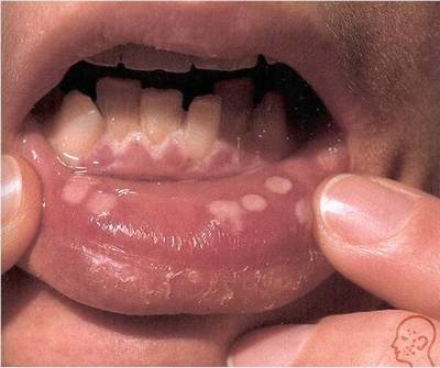
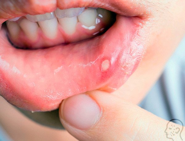
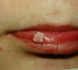
If a blistering rash, each pimple of which is filled with liquid, appears on the inner area of the lip, then a herpetic infection has developed. Herpes simplex virus is the causative agent of the disease. It most often affects the outer surface and skin in the area of the lips and mouth. Herpes on inside lips is one of the most severe forms of the disease. The main manifestation is a bubble on the mucous membrane on the inside of the lip, which brings discomfort, burning, itching, and tingling are felt. The disease is contagious and must be treated. If therapy is refused, the herpetic infection becomes chronic with frequent relapses.
The herpes simplex virus is present in the body of 90% of the world's population. Primary infection occurs in childhood. With good immunity, the virus “sleeps” in the nerve fibers, but is highly viable. Activation occurs under certain conditions, such as:
- physiologically caused decrease in immunity, for example, during pregnancy;
- the presence of an unstable immune system that does not have antibodies to herpes, for example, in infants and infants;
- decreased protective function of the body against the background of acute or chronic internal pathologies;
- a state of severe immunodeficiency, for example, HIV, AIDS, etc.;
- dental diseases;
- Unhealthy Lifestyle;
- poor nutrition.
Since the virus is very contagious, and the main manifestation of its activity is a white bubble on the inside of the lip, there is a high risk of contracting the infection through close contact with a sick person. It happens:
- with kissing, oral sexual contact;
- when using a contaminated toothbrush or lip cosmetics;
- when touching saliva with your hands and transferring its drops to other parts of the body;
- when talking, coughing, sneezing, that is, by airborne droplets.
There are known cases of herpes infection in newborns. Despite the appearance of rashes in the inner area of the lips, infection is possible during childbirth due to the ability of the pathogen to travel along nerve fibers to different parts of the body. Settling on the internal surfaces of the genital organs, it may not manifest itself, but remains infectious.
The causes of recurrence of herpes infection in inner surface lips and mouth are:
- severe hypothermia;
- constant stress, overwork;
- injury to the mouth area;
- frequent colds;
- avitaminosis;
- exhaustion.
Pathogen
Herpes under the lip, often called a “cold,” is caused by a dermatoneurotropic virus. It grows and multiplies in infected nerve fibers and cells. It is also called herpes simplex virus or HSV. The incubation period after infection lasts up to 2 weeks, during which a person is contagious.
Primary infection occurs through contact with a virus carrier. Labial herpes is embedded in the DNA of nerve receptors and remains in a latent (sleeping) form for a longer period of time. At the slightest decrease in the body’s protective function, a relapse of the disease occurs, and then the pathogen penetrates the cells of the mucous membrane of any part of the body, where it begins to actively divide. The process is accompanied by the death of cells, in the place of which small blisters filled with liquid form. Subsequently, they burst with the formation of ulcers. If not treated promptly, the cold takes on a protracted form.
It is impossible to completely get rid of HSV, since its main component is hidden in the DNA of human cells. Therapy can relieve symptoms, reduce the duration of the disease and the risk of relapse.
Stages
The disease occurs in several stages:
- There is a slight tingling and discomfort on the inside of the lip and mouth. Timely treatment allows you to avoid further manifestations of the disease.
- Hyperemia begins with swelling of the inner surface. The person feels a slight itch. Symptoms develop during the first hours of infection activation.
- After 1-2 days, the surface inside the lips and mouth becomes covered with characteristic blisters filled with serous fluid. The size of the formations in diameter varies in the range of 0.2-0.5 cm.
- On the 3rd day, the liquid in the vesicles becomes cloudy, and the white bubbles themselves burst. Weeping wounds form at the site of the rupture. This stage is the most dangerous, as a clear, liquid substrate is released along with a large number of viruses, ready to attack.
- Swelling of regional lymph nodes begins, in particular in the neck.
- Gradually, each wound is covered with a crust, which falls off. The wound begins to scar. At this stage, symptoms such as itching, swelling, and redness subside.
Diagnostics
 The doctor will examine the patient and make a preliminary diagnosis.
 When you first suspect the development of herpes under the lips, you should consult a dermatologist. The doctor will examine the sore spots, make a preliminary diagnosis, determine diagnostic tactics, and, based on the results, choose an appropriate treatment regimen.
The main diagnostic methods are:
- polymerase chain reaction, which takes 30 minutes to complete, accuracy is 70-95%;
- virological tests performed within 20 minutes with an accuracy of 60-85%;
- immunofluorescence reaction requiring 30 minutes to achieve 85-99% accuracy.
Treatment
There is no medicine to completely get rid of the herpes virus, so drugs are used to inhibit its ability to reproduce, which speeds up the healing process.
Common antiviral drugs for the treatment of herpetic rashes on the lips from the inside, they are based on acyclovir. Examples of drugs: Acyclovir, Famciclovir, Valtrex, Virolex, Zovirax. Medicines are presented in different medicinal forms, so they can be used both topically (ointment, gel, cream) and orally (tablets). They need to be lubricated with each new blister or sore.
Paracetamol drugs, such as Paracetamol, Ibuprofen, and antihistamines - Zodak, Fenistil - help relieve accompanying symptoms in the form of pain, itching and reduce inflammation. To maintain and enhance immunity, special interferon correctors with an antiviral effect are prescribed, such as “Kipferon”, “Genferon”, “Viferon”.
The complex treatment regimen includes procedures for rinsing the mouth with analgesics, such as Benzydamine and Chlorhexidine. Lidocaine in gel form will help quickly relieve pain. In some cases, it is necessary to treat herpes with laser therapy, for example, in case of a critical decline in immunity.
An auxiliary measure is the use of traditional medicine. The most popular of them are the following:
- treating sore spots with earwax;
- gluing eggshell film to the blisters;
- processing with a mixture of fresh juice of calendula leaves (1 tbsp.) with 1 tsp. Vaseline;
- frequent compresses of aloe juice;
- fir oil treatment.
Prevention
It is possible to prevent relapses of herpes infection by following simple rules. This:
- conducting healthy image life without smoking and alcohol;
- proper nutrition;
- frequent and long walks in the fresh air;
- daily hardening of the body;
- replenishing the immune system with periodic courses of multivitamins and minerals;
- careful adherence to personal hygiene;
- avoiding close contacts with unfamiliar people (kissing, oral sex, etc.);
- use of personal utensils and hygiene items.
The oral cavity is a kind of mirror of the human body, which reflects the signs infectious diseases, malfunctions of vital systems and organs. Ulcers, plaque, blisters or cracks may form on the mucous membrane.
What disease can cause transparent blisters to appear on the mucous membranes of the mouth? The cause of the pathology can be several ailments.
If you consume too hot liquid or food, a burn to the mucous membrane may occur. There are 3 stages of damage:
- Redness of the tissue appears.
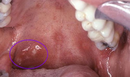
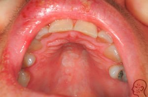
- A watery, transparent bubble appears on the roof of the mouth.
- Dying and rejection of burned tissues.
For mild to moderate burns, the oral cavity should be rinsed with antiseptic solutions; anti-inflammatory gels can be applied to the damaged areas. Before healing, you should avoid eating irritating foods so that the blister does not open and an ulcer forms on the roof of your mouth.
The disease is caused by the herpes virus, and blisters with cloudy liquid appear on the palate, tongue, inside of the lips, cheeks, and there is a burning and itching sensation in the mouth. The nasolabial triangle may also be affected. Before the bubbles appear, patients feel unwell, body temperature rises, mucous membranes hurt and itch, and regional lymph nodes become inflamed. The rashes are usually multiple and can merge into one large lesion.
After some time, the blisters on the oral mucosa spontaneously open. In their place, erosions remain; when an infection occurs, inflammation can develop and ulcers can form. According to the severity, herpetic stomatitis can be mild, moderate and severe.
Treatment is aimed at suppressing the herpes virus. Patients are prescribed regular treatment of the oral cavity with antiseptic agents, and anti-inflammatory and painkillers are applied to the affected areas. Immunomodulators, vitamins and antiviral medications are taken internally.
Dühring's dermatitis herpetiformis
This is a skin disease caused by intestinal dysfunction. Patients develop painful blisters on the skin and mucous membranes of the mouth. External signs very similar to the manifestations of herpes. 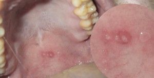 The rashes come in different sizes and types: they can be tense with clear liquid, covered with a crust, or have the form of a papule. Their appearance is preceded by general malaise, chills, itchy skin, burning. The bubbles are most often localized on the hard palate, cheeks, and mouth. The disease is chronic, so relapses occur periodically.
After 3 days, the blisters on the mucous membrane in the mouth open and form erosions. After another 3 days, the wounds heal, leaving an inflamed area or a small scar in their place.
The disease can develop at any age, but most often affects men 30–40 years old. For treatment, sulfone drugs, vitamins, antihistamines, corticosteroids, and a special diet are prescribed.
Vesical vascular syndrome
In people suffering from hypertension or cardiovascular diseases, a dense blister may appear in the mouth on the cheek, soft palate, or tongue. It looks like a single red bubble that stays in the mouth for up to 2 days. This manifestation is called bladder syndrome. The cause of blisters is the rupture of small blood vessels in the mouth when blood pressure rises.
After perforation of the bladder, erosion is formed, which epithelializes after 3–5 days. When infected, suppuration occurs and a deep trophic ulcer is formed.
Vesicovascular syndrome is most often observed in women over 40 years of age. Treatment is carried out under the supervision of a cardiologist.
Erythema multiforme exudative
An inflammatory disease of the mucous membranes and skin is called erythema. The acute course is manifested by the formation of blisters, papules, and blisters in the mouth. The course of the pathology is long-term with the occurrence of periodic  relapses. The rashes are most often localized on the inside of the lips, cheeks, tongue, soft palate, and floor of the mouth.
Before the appearance of blisters, patients complain of general malaise, fever from 37˚ to 38˚, burning in the mouth, aches throughout the body. Afterwards, hyperemic spots appear, in the center of which a bubble filled with serous fluid forms. Painful sensations are constantly present. Patients cannot talk or eat.
The blisters break open after a few days, and in their place erosions form, covered with a fibrous coating. When wounds become infected, inflammation occurs, the ulcers become covered with a yellow-gray coating, which is also found on the teeth and tongue. Regional lymph nodes become inflamed and salivation increases.
An exacerbation lasts 2–3 weeks, healing of erosions occurs in 7–10 days without tissue scarring. Treatment consists of taking disensitizing, anti-inflammatory drugs, and vitamins. Antiseptic treatment of the oral cavity and erosions is carried out locally. Severe forms of erythema are treated in a hospital setting under the supervision of a physician.
Pemphigus
A flaccid transparent bubble has appeared in the mouth, what is it? This may be a manifestation of an autoimmune pathology - pemphigus. It most often affects people over 50 years of age. There are several types of disease:
Pemphigus is a dangerous disease, it can be benign and malignant, and therefore requires immediate treatment from a dermatologist and dentist.
Epidermolysis bullosa
This is a genetic pathology that affects newborn children. The disease has several forms (simple, borderline, dystrophic), the clinical manifestations depend on this. With any of its types, thinning of the skin and mucous membranes is observed; with minor trauma, a transparent bubble with liquid can form in the mouth, on the palate, or in any part of the body.
First, a tense blister filled with cloudy fluid appears on the affected area in the mouth. After opening it, painful erosions and ulcers form, and candidiasis may occur. After healing of deep wounds, the tissues scar and lead to deformation of the mucous membranes and malocclusion.
Pathology can affect anyone internal organ, skin, bones, eyes, hair and nails. Unfortunately, the pathology is incurable.
nashizuby.ru
Possible reasons
Some experts call the oral cavity a mirror that reflects the level of general health of the patient. The appearance of various bubbles on mucous tissues can signal specific pathologies, infectious or non-infectious diseases, acute and chronic ailments:
- endocrine system;
- hematopoiesis;
- heart and blood vessels;
- respiratory organs;
- kidney
In addition, formations in the mouth can be a sign of hypovitaminosis, a reaction to chemotherapy, and even a symptom of syphilis, immunodeficiency, or a cancer process.
Some of them can be identified immediately upon visual inspection, others require more thorough diagnosis.
In any case, the reasons, as well as specific measures of assistance, must be dealt with by the doctor.
But there are also specific diseases that affect only the mucous membranes of the mouth.
Stomatitis and “company”
More often than infectious diseases, pain and inconvenience bring various mucosal injuries– burns from hot food or drinks, scratches from sharp objects (cutlery or the corner of an orthopedic structure, fillings), allergic manifestations due to various substances entering the mouth.
Alas, there are many cases on the “conscience” of viruses, bacteria and fungi, even in childhood. Congenital pathologies also occur.
Stomatitis
An infectious disease that occurs in acute or chronic form. Most often, it is this that causes transparent bubbles in the mouth on the palate. Most common herpetic form of stomatitis, In second place - aphthous type of pathology.
For adequate therapy, it is necessary to know exactly the cause of the disease and the type, since drugs that act on some pathogens have no effect on others. For example, the herpes virus is eliminated by acyclovir and its derivatives, but these drugs are useless for eliminating fungal or bacterial infections.
Congenital epidermolysis
Congenital epidermolysis, also called congenital pemphigus. The disease can be simple or dystrophic. Congenital pemphigus is usually found in early childhood and can accompany the patient throughout all my life. Blistering formations in this pathology form on the palate and tongue, on the inner surface of the cheeks and lips.
Treatment of a simple form is usually symptomatic. In the dystrophic form, in severe cases, corticosteroid therapy is indicated. Nutrition plays an important role in the treatment of pemphigus: it should be nutritious, high-calorie, but salt-free. Anesthetics are used for local treatment of the oral cavity. There are also other types of pemphigus related to autoimmune diseases, which, fortunately, are extremely rare (paraneoplastic, Brazilian and foliate forms).
Hand-foot-mouth syndrome
This disease occurs mainly in childhood and is provoked Coxsackie virus. It is not difficult to recognize: watery transparent bubbles appear in the mouth on the mucous membrane, on the palms and soles. No specific therapy is required, and symptomatic care includes preventing dehydration and relieving fever and pain.
Dühring's disease
Dühring's disease or, in other words, dermatostomatitis herpetiformis. Although the etiology of this pathology has not been identified, it is classified as pemphigus. The disease is accompanied by rashes in the form of spots, blisters and blisters on the skin and mucous membrane of the oral cavity, and on the skin, as a rule, elements of the rash appear more often.
The formations are painful, cause discomfort and itching. Often a secondary infection is associated with the pathology. Therapy is based on drugs from the group sulfonamide. If they have no effect, they resort to hormonal drugs that give quick results. Local assistance includes treating the affected mucosa with disinfectant solutions and pain relief.
Shingles
This disease is also provoked by the herpes virus and is accompanied by the formation of small, painful spots, which, as the disease progresses, turn into blisters.
Note! This disease can only appear in people who have previously had chickenpox. The pathogen does not disappear from the body after chickenpox ends, but “falls asleep” and reminds itself of itself at every opportunity.
And there are many situations in which herpes becomes active again:
- nervous experiences and shocks;
- decreased immune defenses due to prolonged or irrational use of certain medications;
- acute and chronic diseases;
- oncological processes;
- chemotherapy;
- chronic fatigue;
- autoimmune diseases and immunodeficiency.
The disease is contagious Therefore, it is advisable for patients with shingles not to have contact with other people until complete recovery, especially with young children and adults who are not immune to chickenpox.
Treatment includes symptomatic care, prevention of suppuration of blisters through personal hygiene, antiviral therapy with acyclovir and other similar drugs. In severe cases, when shingles is not limited to the skin or oral mucosa and affects the eyes, it is necessary urgent hospitalization.
Herpangina
Herpangina ( do not confuse with sore throat!). The culprit of the disease is coxsackie virus, of which there are many types. The course of the disease is similar to acute bacterial tonsillitis, but there is a significant difference in both symptoms and treatment. In the case of herpangina, the inflamed areas in the mouth become covered with small spots 2-3 days after the first signs, which very quickly turn into blisters. After another 2–4 days, these elements burst, leaving behind small wounds. As a rule, on the 6th–7th day of illness, all phenomena disappear and recovery occurs.
Specific therapy for uncomplicated herpangina does not require. Drugs are prescribed for symptomatic relief, rinses are used, a gentle regimen and temporary isolation at home are recommended. In case of severe illness or secondary infections, the patient is immediately sent to the hospital under medical supervision.
In addition to these fairly common reasons, bubbles, spots and blisters on the roof of the mouth can also appear due to other diseases.
Is it possible to be treated at home?
Undoubtedly, since most of the identified diseases do not require stay in medical institutions. However, the cause of the disease and methods of influencing the “culprit” must be determined by the doctor. The task of adult patients and especially parents is not to wonder whether it is a hard bubble popping up on the roof of their mouth or a scattering of small bubbles appearing on the tongue, but to quickly get ready to see a specialist.
As for traditional medicine recipes, which are sent in large quantities to in social networks and are advised by numerous acquaintances, then patients should be aware: the use of such methods for diseases that go away on their own is useless, but for the most part safe. And for diseases that require specific intervention medicines and even hospitalization, the folk recipe is ineffective and dangerous, primarily due to the loss of time.
vashyzuby.ru
Causes of oral herpes
Herpes in the mouth appears as a result of human infection with the herpes simplex virus type 1 or 2. Herpes simplex penetrates the nerve plexus and waits for favorable circumstances to occur, after which it makes its way through the nerve axons to the skin and provokes its inflammation.
Various factors contribute to the activation of herpes on the oral mucosa:
- Stress.
- Avitaminosis.
- Weakness of immunity.
- Frequent colds.
- Surgical intervention.
- Oncological pathologies.
- Increased physical activity.
- Chemotherapy and antibiotic treatment.
- Exposure to low or high temperatures.
- Hormonal fluctuations in women during menstrual days.
It is easy to catch a herpes infection through kissing, unprotected sexual contact, incl. and with oral sex, as well as when using common hygiene items. The risk of infection increases when the partner goes through an acute stage of the disease or has specific rashes on the lips or oral mucosa.
In carriers of the herpes virus, the disease may be asymptomatic. However, such people become a source of infection, because the pathogen is present in their saliva, blood, and tears. From here follow the routes of infection with herpes - sexual, contact, airborne, transfusion and transplacental.
Signs of oral herpes
The primary symptoms of herpes in the mouth include tingling, stinging and itching sensations. Next, slight swelling and redness occurs. Eating is difficult due to pain.

At the next stage, bubbles form, which after 3 days burst and turn into painful yellow erosions. The oral cavity seems dry. Gradually, the ulcers become overgrown with dense crusts that are prone to bleeding. After 10–14 days, the lesions heal without tissue scarring.
Doctors distinguish three degrees of severity of herpes in the mouth:
- Mild, asymptomatic. But if you carefully examine the oral cavity, you will notice swelling of the delicate mucous membrane and small wounds. Slight fluctuations in body temperature are possible.
- Average. This form is characterized by pronounced symptoms with changes in blood composition (they are determined by tests). There is no point in delaying the treatment of moderate oral herpes, because... With timely treatment, the problem is resolved faster.
- Heavy. The form is characterized by a sharp deterioration in health and an abundance of rashes on the lips and inside the oral cavity. The body temperature jumps to 40°C, the lymph nodes of the cervical and submandibular zone become inflamed, a blood test shows an increase in ESR.
To clarify the diagnosis, the doctor suggests taking a smear for analysis or doing a biopsy of the herpetic element. This is necessary when the patient is in serious condition, or difficulties arise when making a diagnosis visually.
The difference between herpes and other diseases
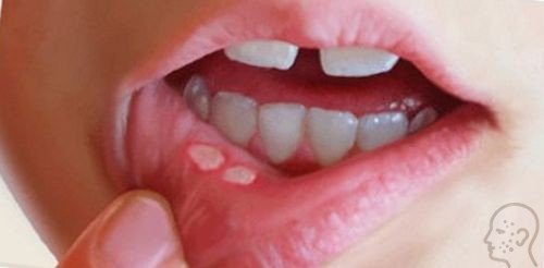
People often perceive herpetic rashes in the mouth as signs of stomatitis. Common to both pathologies are painful ulcers that resolve on their own in 1 to 2 weeks.
The following conditions help differentiate herpes from stomatitis:
- Herpes simplex affects the area of the mouth adjacent to the bones. Signs of stomatitis are found on the inner surface of the lips, cheeks and throat.
- Herpes manifests itself as vesicles, after opening which leave ulcers. With stomatitis, the oral cavity ulcerates immediately.
- The herpes virus is localized in one area. Stomatitis affects different places.
Diagnosing herpes in the mouth is not difficult, look at the photo and you will see that it looks the same in adults. The only thing is that the bubbles can be of different sizes.


Therapeutic measures for herpes in the mouth
The correct treatment for herpes in the mouth in adults is considered to be one that is based on integrated approach. This is the use of medications, the use of vitamins and diet. In the acute period of a herpetic infection, it is important to maintain water balance in the body and drink plenty of clean liquids.
The first signs of the disease must be combated with antiviral drugs:
- Acyclovir.
- Zovirax.
- Megosin.
- Famvir.
- Valtrex.
- Diolin.
- Holisal.
- Solcoseryl.
In mild forms, oral herpes is treated with topical medications. In advanced cases, take systemic tablets.
Mouth rinses are carried out with salt water, a solution of Furacilin, Miramistin or Chlorphilipt. The pain is relieved with Kalgel (contains lidocaine).
For immune stimulation, patients are simultaneously prescribed special drugs - Decaris, Imudon, Histaglobulin. Multivitamins and lectraves – rosehip, viburnum, echinacea – help increase the body’s resistance to any viruses. Antipyretics are indicated only for significant hyperthermia.
In the presence of purulent ulcers, antibiotics are added to the course:
- Biseptol.
- Amoxicillin.
- Ceftriaxone.
Folk options for fighting herpes
What else, besides medications, can be used to treat herpes in the mouth of an adult patient? Doctors do not insist on using folk remedies, but do not prohibit them, because some techniques turn out to be quite effective.

- Aloe. Oral intake of fresh juice squeezed from the leaves. Lubricating the affected mucous membrane with juice.
- Fir oil. Used to treat ulcers. The interval between procedures is 3 hours.
- Sea buckthorn oil. It is used like fir.
- Raisin. Dry grapes are cut in half and rubbed on the wounds 3 - 4 rubles. in a day.
- Chamomile, lemon balm, wormwood. The plants are infused for half an hour in boiling water and filtered. Use steam to rinse your mouth.
- Ice. Cubes of frozen water are applied to the corners of the lips affected by herpes.
- Alcohol. Viral lesions on the lips are wiped with medical alcohol.
Diet
While treating herpes in the mouth at home, you need to switch to a gentle diet that will ease the pain and help the inflammation subside. The patient is allowed to eat only warm food. These can be soups, broths, milk porridges.
For herpes, it is useful to eat foods enriched with lysine and arginine:
- Cottage cheese.
- Milk.
- Butter.
Prevention of oral herpes consists of maintaining immunity. To prevent the virus from recurring, it is important to lead a healthy lifestyle, eat with an emphasis on fortified foods, give up bad habits and not engage in intimate relationships with unverified partners.
Video:
P.S. Self-medication, despite our recommendations, is not worth doing. Therapy will be effective only after a thorough examination by a specialist. If you try to treat oral herpes on your own, it may complicate the course of the disease. It will be more difficult to suppress the activity of the pathogen.
kozhnyi.ru
Features of herpes in the mouth
Let us immediately determine that type 1 herpes is present in the body of each of us, but the infection becomes active only during a cold or weakened immune system. That’s why people call the disease a “cold.”

Often the blister appears inside the cheek, on the lips, tonsils or gums. By appearance looks like a watery bubble, sometimes it is a series of small blood bubbles down the throat.
Let us highlight that the disease develops more often in children under five years of age; previously, they get sick less often. Because the immunity received from parents works.
Herpes on the inside of the lip may be:
- Acute When infection with the herpes virus occurs, about 80% of patients in large groups suffer from it;
- Chronic, when blisters pop up periodically, at a time when the immune system is weakened.
If you are faced with the first form of herpes, then you need to start emergency treatment, but if you suffer from the second form, then it is important to periodically carry out prevention.
Let us highlight the forms of development of the disease:
- Lightweight when bubbles appear in the mouth unnoticed, the temperature remains normal, but swelling in the oral cavity is visible. Then the entire area behind the lower lip or cheek is affected in the form of blisters. Adults are less susceptible to this phenomenon than children;
- Average, when the symptoms appear more clearly, but with timely treatment you can get rid of the disease completely;
- Heavy, when herpes spreads not only to the lips, but also to the gums and cheeks. Bubbles appear on the palate unexpectedly, and if left untreated, marks and scars may appear.
Particularly attentive to the appearance of herpes in the mouth should be parents of young children, because often the disease is diagnosed too late, when it is in severe form.

Herpes on the oral mucosa - video
Herpes in the mouth: causes of appearance
We have determined when the blister appeared and what it is, but it is important to understand why this happens.
This is how the virus spreads through airborne droplets, namely:
- Through kisses and other close contact with the patient;
- Insufficient hygiene.
After entering the body, the herpes virus moves to the nerve endings, where it can remain in a passive state for decades. At the time of a cold or general loss of strength, the virus moves into the oral cavity, causing inflammation.

 For activation of herpes in the oral cavity to occur, it is enough:
- Stress and strain;
- Pain syndrome;
- Operations;
- Colds;
- Increase in temperature;
- Influence of environmental factors;
- Menses;
- Autoimmune diseases;
It happens that white watery rashes form and open within 10-12 days after activation. But it is important to distinguish between the symptoms of stomatitis, which forms on the inside of the cheek when the herpes is located closer to the gums and lips.
Also, stomatitis does not have an exact location, when the herpes blisters are located in one area.
Herpes in the mouth - what it looks like: photo
Manifestations
But the most significant manifestations are:
- The appearance of burning and tingling in places where herpes is localized; if the lymph nodes are affected, aching pain is felt;
- The gums and mucous membrane of the mouth change color, darken, and swelling begins. Saliva becomes more viscous, and watery blisters may bleed when pressed on them;
- There is a rash throughout the oral cavity, which has a transparent tint, but is filled with liquid;
- Sores that burst may ooze yellow fluid and become crusty. Sometimes there are cracks and small scars;
- After the wounds heal, the gums continue to bleed, and the swelling remains mild.
You need to understand that the first time herpes will look like stomatitis, which often happens to children, but the second time the herpes will be localized on the lips and gums.
![]()
Below we will look at the differences between herpes and its symptoms, depending on the location in the mouth:
| Localization location | Peculiarities |
|---|---|
| Herpes on gums | Lasts about a week, looks like a rash on the mucous membrane, pain and bleeding of the gums may be felt. The wounds are covered with a yellow coating, the gums are covered with pus, sometimes the inside of the lip is affected, and after healing there are no wounds left. |
| On the inside of the lips | There is a burning sensation and itching, the affected area swells and turns red. Several clear bubbles containing liquid form. After a couple of days, a small ulcer with a crust may appear; after a week, the blister peels off, but cracks with blood remain. |
| Herpes on the palate | There may be several wounds in different parts of the palate or a rash on the tonsils, which is a severe form. There is almost no swelling, but the wounds, after they burst, leave scars and marks. |
Important prevent the development of herpes on the tonsils and tonsils, because then the blisters will have the appearance of erosion, turning into ulcers with tissue necrosis.
Then the general symptoms may be joined by:
- Pain when swallowing;
- Allergy;
- Candidiasis;
- Difficulty breathing;
- Development of pathologies of the digestive tract.
Why do mouth ulcers appear and how to treat them?
Therapy
Therapy to get rid of rashes on the palate must be complete, usually it involves:
- Bed rest for severe cases;
- A diet with limited sweet, salty and spicy foods;
- Maintaining water balance;
- Use of medications;
- Taking antipyretics;
- Use of antiviral agents;
- Reception vitamin complexes and ascorbic acid.
Medications that should be used include::
- Interferon;
- Chlorhexidine bigluconate;
- Riodoxol or oxolinic ointment;
- Infusion of calendula or rosehip;
- Megosin;
- Holisal.

Note that Cholisal for herpes on the lips has a general effect, stimulates the immune system, helps with lungs colds. But you can take it only as prescribed by a doctor.
If the herpes is not located on the lips, but on the inside of the cheeks, in the oral cavity, it is better to use not ointment or oil, but tablets that are not washed off with saliva. At the same time, it is recommended to use antiseptics and dental solutions to get rid of the bacterial infection.
Can be used to relieve pain and eliminate general symptoms.:
- Tantum verde with analogues;
- Special pastes;
- Decoctions or ointments based on chamomile, calendula or St. John's wort.
Note that It is better not to use analgin or aspirin in this situation, since there is a high probability of complications and adverse reactions.
Traditional therapy
If herpes has just appeared on the lips or the inside of the cheeks, then you can resort to traditional medicine methods, which include:
- Applying toothpaste to a blister that has just appeared, when bacteria will not multiply and the wound will begin to dry out;
- Sprinkle a thin layer of table salt on your lip several times a day;
- Make poultices or compresses based on chopped garlic and grated apple, mix them in equal proportions, one teaspoon at a time;
- Make a compress from cooled black tea, applying gauze to the affected area for 20 minutes;
- You can lubricate the bubbles with valocordin or sage oil;
- Prepare an ointment based on walnuts and honey, which are mixed in equal proportions and applied to the blister;

Treatment of herpes in the mouth at home: video
Prevention of herpes
It is impossible to completely exclude the development of herpes in the inside of the mouth, because about 90% of the population is susceptible to it globe. Moreover, the disease occurs in an asymptomatic form, so there is no point in talking about timely treatment. You can only carry out prevention and eliminate the occurrence of relapses.
This is done by following these rules:
- Maintaining a healthy lifestyle;
- Rejection of bad habits;
- Maintaining immunity;
- Diet;
- Staying outdoors every day;
- Avoiding stress;
- Undertaking preventive examinations and treating colds.

Also avoid close contact with people with herpes for two weeks., this will prevent the virus from activating in your body. To do this, it is enough to avoid getting saliva or blood through dishes, a toothbrush and kisses.
Conclusion
As it became clear, it is impossible to completely avoid the appearance of herpes, and we often do not notice relapses at all, because the disease occurs without pronounced symptoms.
But by triggering inflammation and the development of rashes, you can encounter damage to the tonsils and respiratory system, the development of pathologies of the digestive tract, necrosis, and ulcerative damage to body tissues. Such problems have serious consequences, which will take a month to get rid of.
To prevent this from happening, be attentive to your well-being, do a weekly examination of your child’s oral cavity, avoid contact with sick people, maintain immunity and treat colds in a timely manner.
Only then will a minor problem like herpes not become a problem that torments you throughout your life. And following the rules of prevention will allow you to avoid any relapses, discomfort, the appearance of wounds and scars in the mouth. And this will give health not only to the gums, but also to the tonsils, teeth, and tonsils.
zdorovkozha.com
Characteristics of a blood bubble on the oral mucosa
 The mucous membrane protects the entire body from negative influence environment, from harmful microorganisms, various types of pollution, and also has a fairly high level of regeneration. If blood blisters regularly appear on the oral mucosa, then you should take this signal seriously and take action.
A bloody ball in the mouth is a hematoma (bruise), which is characterized by the accumulation of blood in a certain place in the oral cavity. The appearance of bloody blisters is a kind of hemorrhage that occurs due to trauma to the capillaries and thin vessels of the mucous membrane.
A blister on the mucous membrane may contain clear serous fluid without the presence of blood. This means that the vessels were not damaged and the resulting wound is superficial. Such blisters on the mucous membrane heal much faster. The presence of blood in the bladder indicates a deep injury and a longer period of healing and blood resorption.
The main causes of a blood blister
 The general condition and integrity of the oral mucosa usually indicates the level of health of the body. Often through research appearance oral mucosa and blisters, the doctor makes a final diagnosis. After all, the symptoms of most infectious, bacterial, chronic, and acute processes that occur in the body are associated with changes in the integrity and color of the oral mucosa. Therefore, it is important to understand the main reasons that cause blood blisters to appear in the mouth.
Blood blisters are distinguished by the place of their occurrence - on the tongue, under the tongue, on the cheek. They can occur as a result of injury or be a signal of the presence of a serious disease in the body. Multiple blood blisters on the oral mucosa occur with stomatitis, diseases of the gastrointestinal tract, and disturbances in the functioning of the endocrine system.
The cause of the sudden appearance of a blood bubble in the mouth is damage to the mucous membrane.
There are the following types of injuries to the oral cavity:
- mechanical injury. The cause may be various objects, solid food, biting the cheek;
- chemical injury. It occurs due to the consumption of spicy, salty foods, and exposure to chemicals on the mucous membrane. This irritates the delicate oral mucosa and causes injury;
- thermal injuries. Their appearance is provoked by too cold or hot food or drinks.
The mechanism of formation of a blood bubble on the oral mucosa
 Bloody blisters in the mouth in most cases are not life-threatening. They are formed as a result of mechanical damage to the mucous membrane. When microtrauma occurs, harmful microorganisms attack the damaged area.
After this, a number of responses are activated in the human body:
- The immune system is activated. Monocytes and leukocytes, as well as macrophages, instantly arrive at the damaged area, attacking the harmful pathogen and quickly destroying it.
- Immune cells die. This is a signal for other cells and substances are released in the affected area that are mediators of inflammation of the mucous membrane - serotonin, histamine and bradykinin.
- These substances cause a strong spasm of the circulatory system and the outflow of blood is hampered. After the spasm is relieved, all accumulated blood immediately flows to the site of inflammation. It moves at high speed and under pressure. A detachment of the mucous membrane occurs in the mouth, and a bloody blister appears.
Treatment of bloody blisters in the mouth
 A blood blister in the mouth is only part of the body’s defense reaction and goes away on its own within a week. If this does not happen, then you should consult a doctor to rule out serious diseases of the body and neoplasms. He will be able to make an accurate diagnosis after a thorough examination, studying the data of clinical tests and histology. After this, the doctor will prescribe the correct treatment.
The process of treating a blood bladder in the oral cavity is closely related to the cause of its appearance and therefore treatment depends on several important factors:
- volume of surface damage;
- degree of filling with serous fluid;
- the nature of the contents of the blood bladder;
- location.
The volume and nature of the damaged surface is important when prescribing treatment for a bloody blister in the oral cavity. After all, the larger the volume of the blood bladder, the worse it heals and resolves. Treatment of a large bladder with blood can develop from conservative treatment into surgical intervention. Small blood blisters resolve quickly and do not require special treatment.
A blood blister on the oral mucosa must be carefully examined to exclude hemangioma and vascular tumor. A doctor can do this when examining the oral cavity. Hemangioma is sometimes left without much treatment if it does not grow. At intensive growth it should be removed surgically.
Many bloody blisters in the mouth can be associated with syphilis, sometimes pemphigus. Small red blisters on, under, or on the side of the tongue may indicate the presence of glossitis, an inflammation of the surface of the tongue caused by harmful microorganisms. Treatment will consist of treating and rinsing the mouth with antiseptic solutions and eliminating the disease, which has become main reason the appearance of blood blisters.
 It is not necessary to treat a bloody blister in the mouth if it is isolated and does not bother the person. If it interferes, the doctor performs a puncture after a thorough examination and diagnosis.
To strengthen the walls of blood vessels and the immune system, vitamins E, A, C, K, B vitamins, and multivitamin complexes are prescribed.
The appearance of bloody blisters in the mouth indicates an oral injury or is a symptom of a disease in the body. Install the real reason Only a doctor can prescribe this education and prescribe effective treatment. If you seek qualified help in time, this disease will not cause discomfort and will not lead to serious consequences.
Dermatology, the study of skin diseases, is of particular interest to dentists not only because many skin diseases also involve the oral mucosa, but because oral lesions are often the primary manifestations of skin diseases.
It is especially important for the dentist to know that in many dermatological diseases, lesions of the oral mucosa are not only a concomitant symptom of a skin disease, but often precede skin rashes, and the dentist can often make a dermatological diagnosis before skin lesions appear.
Pemphigus Definition
Pemphigus is a serious chronic skin disease characterized by the formation of intraepithelial blisters containing fluid.
Etiology is still unknown, although there are numerous attempts to link the occurrence of the disease with microorganisms, viruses, and metabolic disorders.
The disease is based on autoimmune mechanisms, since in these patients intercellular antibodies can be detected in the epithelium of the skin and mucous membranes, and circulating antibodies to the epithelium are found in the blood, although the reason for the increased sensitivity to their own epithelial tissues is unknown.
The name pemphigus (“pemphigus”) was first used by Hippocrates (460 - 370 AD) to designate a severe fever accompanied by the appearance of blisters on the skin. The first description of pemphigus vulgaris belongs to the Swiss Koenig (1681). V. P. Nikolsky (1896) made a great contribution to the study of pemphigus. Specific gravity This disease among all other diseases ranges from 0.7 to 1%, and women aged 35 to 65 years are more often affected. Children rarely get sick. The familial nature of this pathology has not been confirmed.
Pemphigus classification
True acantholytic pemphigus
- vulgar
- vegetative
- leaf-shaped
- seborrheic (erythematous)
Nonacantholytic pemphigus
- actual non-acantholytic pemphigus (bullous pemphigoid)
- mucosynechial atrophying bullous dermatitis (pemphigus of the eyes, cicatricial pemphigoid)
- benign non-acantholytic pemphigus of the oral mucosa only
Acantholytic pemphigus
All clinical varieties of true pemphigus are characterized by the presence of acantholysis, which consists in the melting of intercellular connections, degenerative changes in the nuclei and loss of part of the protoplasm; as a result, there is a disruption of communication not only between the cells of the Malpighian layer, but also between the layers of the epidermis.
Such so-called acantholytic cells are easily detected in a fingerprint smear taken from the bottom of the bubble or the surface of the erosion. Each clinical stage corresponds to a specific cytological picture.
The blisters are formed due to acantholysis within the epidermis.
Pemphigus vulgaris on the skin:
Pemphigus vulgaris is more common than other forms. The disease begins acutely. Flaccid blisters appear on apparently unchanged skin. On the body, blisters appear in the folds, on the limbs, on the scalp, under the mammary glands.
Sometimes the bubbles are the size of a chicken egg, their contents are yellowish, translucent, large bubbles take the shape of a pear - "pear symptom" . This symptom is absent in other bullous dermatitis.
Evolution of the Primary Elements
Bubbles easily turn into erosions. Gradually, the erosions increase and, merging with each other, capture new foci, forming continuous lesions. They become covered with crusts, under which epithelialization does not occur.
Skin lesions are combined with fever, depression, psychosis, gastrointestinal dysfunction, etc. All this leads to cachexia, which is fatal.
Phases of the course of true pemphigus (N. D. Sheklakov)
In the first phase (initial) rashes on the oral mucosa look like single blisters and erosions; blisters and epithelial erosions are observed on the skin, which leave behind pigmentation. The general condition of the patient is satisfactory.
Second phase (generalized) characterized by many merging erosions on the mucous membrane and skin, Nikolsky’s symptom is positive. Skin lesions are extensive, fever and intoxication are observed. Patients often die.
In the third phase Epithelization occurs under the influence of treatment with corticosteroids. Nikolsky's symptom is difficult to cause; erosions on the skin become epithelialized. When the patient's condition improves, Nikolsky's symptom becomes negative.
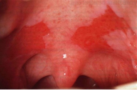
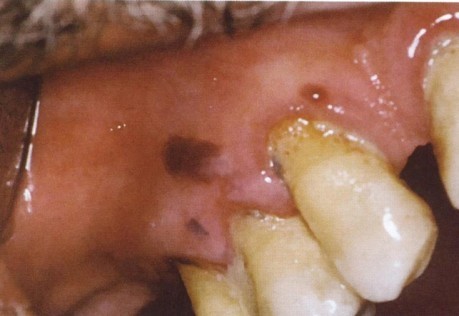
In most cases, the process begins with the oral mucosa.
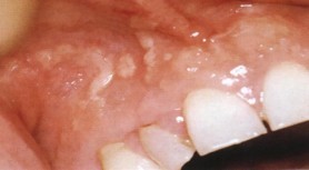
Bubbles are located in the mouth, on the mucous membrane of the cheeks, on the gums, tongue, and pharynx. Opening up, they turn into erosions, bordered by fragments of the epidermis, and when merging, they form continuous foci. Similar rashes can occur in the pharynx and esophagus. When the mucous membrane is affected, there is abundant salivation and there may be an unpleasant odor.
Clinical manifestations of pemphigus
Lesions of the oral mucosa are similar to skin manifestations, although due to the structural features of the mucosal epithelium - the absence of a stratum corneum - an intact bladder in the mouth is extremely rare, since it tends to rupture with the formation of erosion before its complete formation.
Erosion can be extremely painful, leading to the inability to eat. Erosions have uneven edges, their surface is often covered with fibrinous white or blood-stained plaque.
Pemphigus vulgaris is characterized by rapid formation of blisters, varying in size from several millimeters to several centimeters, on apparently healthy mucosa, without signs of inflammation. These blisters have a thin covering and contain clear exudate, which can soon become hemorrhagic or purulent. When the lining of the bladder is opened, the eroded surface is exposed.

Spread to the red border with the formation of hemorrhagic crusts is quite common. There is not a single zone resistant to the disease.

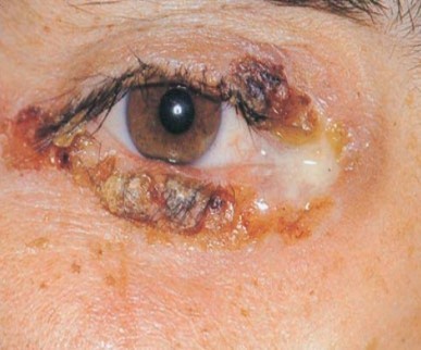
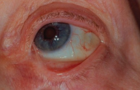
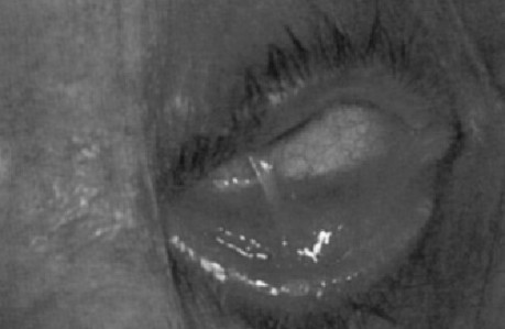
Pemphigus vulgaris, eye damage:
Nikolsky's symptom:
Nikolsky's symptom is a peripheral expansion of erosion when pulling on the remnants of the bladder cover.

A characteristic symptom of true pemphigus is Nikolsky's symptom - the appearance of a bubble or the formation of erosion when rubbing apparently unaffected skin or mucous membrane. Occurs as a result of disruption of communication between spinous epithelial cells and intercellular edema.
Pemphigus foliaceus
Characterized by rapid opening of primary flaccid intraepithelial blisters and drying of the exudate with the formation of massive layered crusts resembling eczema or exfoliative dermatitis. Characteristic is the repeated formation of blisters under the crusts.
The disease can develop from other forms of pemphigus or occur primarily as pemphigus foliaceus.
This is a relatively mild form of pemphigus , which mainly occurs in older people. There is an endemic form of pemphigus foliaceus, found in tropical areas, known as Brazilian pemphigus, which occurs in children and often in members of the same family.
Pemphigus vegetans
It is much less common than vulgar. Flaccid blisters, smaller than in pemphigus vulgaris, are eroded and vegetations form on the surface of some of them. These vegetations are covered with purulent exudate and surrounded by an area of inflammation. The vegetative form most often occurs on the nose, in the corners of the mouth, axillary and anogenital areas, and often resembles condylomas lata, characteristic of secondary recurrent syphilis. The course of the disease is the same as with pemphigus vulgaris, but the vegetative form is characterized by longer remissions.
Erythematous pemphigus
It was first described in 1926. This type of pemphigus is characterized the formation of flaccid intraepithelial blisters with a thin covering and erythematous-squamous plaques, reminiscent of seborrheic dermatitis or lupus erythematosus. The face is most often affected, and the lesion has the shape of a butterfly with hyperkeratosis and blisters. The process also sometimes spreads to the body, where it develops in the form of separate lesions. The disease can drag on for years. Periods of remission after exacerbation are common, but in many patients the disease ultimately progresses to pemphigus vulgaris or pemphigus foliaceus. Despite the individual clinical characteristics of these forms of pemphigus, there are a number of common features that are common to them, which constitute the main essence of the diseases. First of all, the primary element of damage to any type of pemphigus is always an intraepithelial bubble, even if in the later stages of the disease there may be various manifestations in the form of crusts and papillomatous growths. Secondly, skin lesions occur sooner or later, although the oral mucosa can often be affected primarily, with the exception of pemphigus foliaceus and erythematous pemphigus.
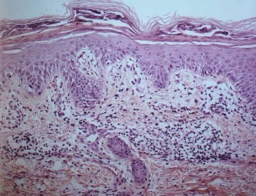
Histological picture of true pemphigus

Pemphigus is characterized by the presence of a bubble located intraepithelially. Histologically, the suprabasilar cleft is clearly defined above the layer of basal cells. At an early stage, edema weakens the junctions of epithelial cells and interepithelial connections are destroyed. This process is called acantholysis. As a result of the destruction of connections between epithelial cells, groups of epithelial cells and individual cells are determined.
Histological picture of pemphigus foliaceus:
Pemphigus foliaceus is characterized by the presence of a bladder located intraepithelially, the covering of which is represented by layers of hyperkeratosis.
Tzanck cells – acantholytic cells
A cytological examination of fingerprint smears from the surface of fresh erosions reveals Tzanck cells lying freely in the intercellular space with a giant hyperchromatically colored nucleus. Such scrapings form the basis for a quick laboratory diagnosis of pemphigus - the Tzanck test.

Interestingly, the bladder fluid contains relatively few inflammatory cells - lymphocytes and polymorphonuclear leukocytes. There are few of them in the underlying connective tissue, which is characteristic feature malignant pemphigus, unlike other vesical lesions, where inflammation is severe. However, when a secondary infection occurs, this picture is quickly masked.
Immunofluorescent methods
Immunofluorescent methods are important in establishing the diagnosis of pemphigus, especially when clinical and cytological findings are inconclusive.
Indirect immunofluorescence is also used to confirm the diagnosis of pemphigus. It is performed by incubating normal animal or human mucosa with serum from a patient suspected of having pemphigus, supplemented with antiglobin-linked fluorescein. A positive tissue reaction indicates the presence of circulating antibodies. A positive indirect reaction in 100% of cases indicates a disease.

Direct immunofluorescence is used to detect immunoglobulins, predominantly IgG, sometimes in combination with IgM and IgA, together with the C3 complement fraction, in the intercellular spaces in the affected oral epithelium, but more often in the unaffected epithelium located next to the lesion. This test is carried out by incubating a biopsy of the mucous membrane of a patient with suspected pemphigus (either frozen samples, or fixed in a special fixative) with antiglobulin combined with fluorescein.
Nonacantholytic pemphigus (pemphigoid)
With non-acantholytic pemphigus, blisters form due to an inflammatory process. The blisters form subepithelially.
Bullous pemphigoid
Bullous pemphigoid is significantly different from pemphigus vulgaris, but has many similarities with ocular pemphigus. Some authors believe that these are simply different variants of the same disease.
Bullous pemphigoid is a disease predominantly of older people, affecting people over 50 years of age.
In approximately 10% of patients, the rash begins in the oral cavity. In the pathogenesis of the disease, autoimmune mechanisms aimed at basement membrane antigens have been proven. Consequently, blisters arise under the epithelium with the involvement of the underlying mucosa, in which signs of inflammation are revealed to varying degrees.
Skin lesions begin as generalized nonspecific eruptions predominantly on the thighs, which appear as urticarial or eczematous eruptions, lasting several weeks or months before becoming vesiculobullous lesions. These bullous lesions have relatively thick walls and may remain intact for several days. If the integrity of the bladder cover is damaged, an eroded surface is exposed. Erosion heals quite quickly.

In the oral cavity, blisters are much less common than with pemphigus vulgaris and with pemphigus of the eyes. On the edematous and hyperemic mucosa, bubbles appear, measuring from 0.5 to 2 cm, with a tense tire, with serous, less often hemorrhagic, contents.
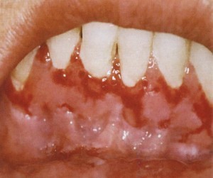
Gingival involvement is typical. Most of the gum is involved. The gum is sharply hyperemic, sharply painful, and desquamation is noted as a result of even minor trauma. However, such lesions can also occur in other areas of the mucosa.
Bullous pemphigoid:


Histological picture of pemphigoid:

Bubbles arise under the epithelium with the involvement of the underlying mucosa, with signs of inflammation to varying degrees.
Mucosynechial atrophying bullous dermatitis
Mucosynechial atrophying bullous dermatitis (pemphigus of the eye, pemphigus of the conjunctiva, cicatricial pemphigoid) is observed mainly in older people.
Blisters form, followed by the formation of scars, adhesions and atrophic areas appear on the skin and mucous membranes of the eyes, mouth, nose, pharynx and genitals. The disease lasts for many years.
In benign pemphigus, the blisters are located subepidermally (there is no acantholysis).

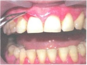
Desquamative gingivitis:

Benign nonacantholytic pemphigus of the oral mucosa only
It is characterized by the appearance of subepithelial (without acantholysis) blisters only on the oral mucosa. Mostly women over 40 years of age are affected. The disease is prone to self-occurring remissions.

In all forms of non-acantholytic pemphigus, Nikolsky's symptom is absent, but detachment of the entire epidermis can be observed at a distance of 3-5 mm from the lesion.
Dühring's dermatitis herpetiformis

With Dühring's dermatitis, a polymorphic rash appears, accompanied by itching and burning. General condition is satisfactory. Mucous membranes are rarely affected. Nikolsky's symptom is negative. Eosinophilia is observed in the contents of the blisters and in the blood. The disease lasts for years, but the prognosis is favorable.
Herpetiform stomatitis:

Differential diagnosis
Erythema multiforme exudative
In isolated lesions of the oral cavity, it can be mistaken for pemphigus. The disease begins acutely, is accompanied by fever and lasts 10-14 days. General symptoms, fever, sore throat, and joint pain may be observed. The blisters are surrounded by an erythematous rim, tense, Nikolsky's sign is negative, acantholytic cells are absent. The skin may have erythematous vesiculobullous forms, when blisters appear on an erythematous base. The contents of the bubbles are transparent, quickly drying into a crust, after which a pigmented spot remains.


Shingles:

The rash with herpes zoster is one-sided, the blisters are located in groups, in the oral cavity - along the II and III pairs of nerves, accompanied by neuralgic pain. Nikolsky's symptom is negative.
Chronic recurrent aphthous stomatitis:

In chronic aphthous stomatitis, aphthae appear on the oral mucosa, erosions are surrounded by an erythematous rim with a yellowish-white coating, are painful, and last 9–13 days.
Pemphigus vegetans must be differentiated from condylomas lata (during the secondary period of syphilis), which can be localized in the corners of the mouth.
Tests for Treponema pallidum, serological and cytological data help to make a correct diagnosis.
![]()